Figure 7. PEDF gene transfection protects against the loss of RGC in DBA/2J mouse retina and delays vision loss. A: Retina flat whole-mount shows extensive GFP labeling 9 months after transfection with AAV-GFP (at 11 months of age). B: Retina cross section shows GFP labeling in ganglion cell layer and in inner nuclear layer. C: Western blot for PEDF in retina from DBA/2J mice 9 months after AAV-PEDF transfection. D: Protection of RGC in DBA/2J mice retina 9 months after transfection with AAV-PEDF. E: RGC loss in retina 9 months after transfection with AAV-GFP. Insets show higher magnification. F: Visual acuity in mock transfected, AAV-GFP, and AAV-PEDF transfected DBA/2J mice, in comparison to C57BL/6J mice. Acuity
decreased progressively beginning at 4 months of age (2 months post transfection) in control transfected and AAV-GFP treated
mice, but was maintained to age 11 months in AAV-PEDF transfected mice. Abbreviations: retinal ganglion cell (RGC) layer;
inner nuclear layer (INL).
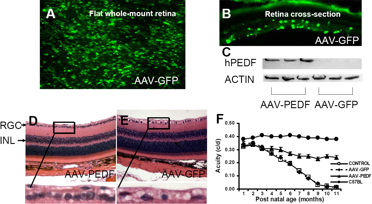
 Figure 7 of
Zhou, Mol Vis 2009; 15:438-450.
Figure 7 of
Zhou, Mol Vis 2009; 15:438-450.