Figure 3. Immunofluorescent localization of PEDF in retina from DBA/2J and C57BL/6J mice. A: PEDF immunoreactivity localizes to the RGC and RPE layers in DBA/2J mice at 1 month old. B: Decreased PEDF expression in the RGC layer of DBA/2J mice at 6 months, with residual staining in the RPE. C: In C56BL/6J mice, PEDF is localized mainly in the RGC and RPE layers. Insets show higher magnification.
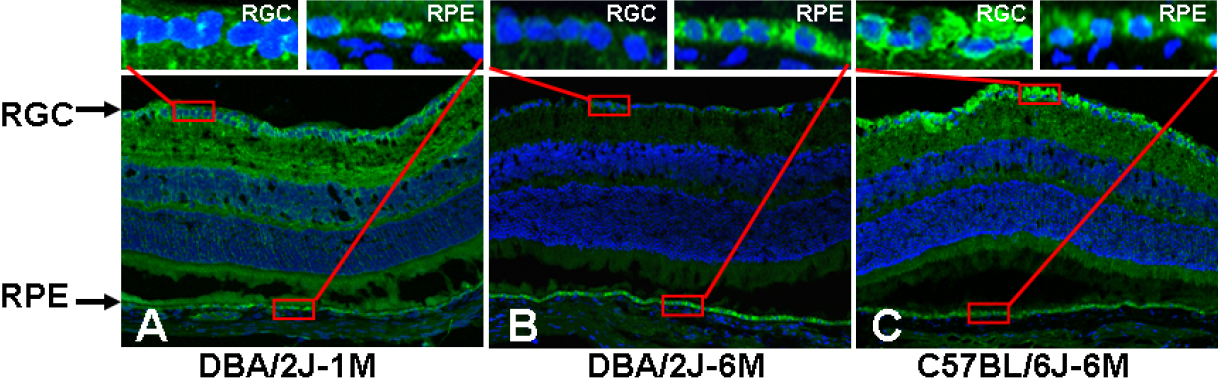
 Figure 3 of
Zhou, Mol Vis 2009; 15:438-450.
Figure 3 of
Zhou, Mol Vis 2009; 15:438-450.