Figure 1. Four novel CYP1B1 mutations observed in Chinese patients with PCG. A: Result of the restriction fragment length polymorphism (RFLP) analysis of the four mutations. “M”: marker; “P”: PCR patient
products; “+”: restriction fragments from patients; “−”: restriction fragments from controls. The size of the fragments is
indicated by an arrowhead. B: Sequencing of the four novel mutations and normal controls are shown. As for the deletions, sequencing of the cloning was
shown. The exact mutation was labeled under each sequence according to the nomenclature recommended by HGVS. C: Pictures of the patient with I471S show the larger cornea and bigger eyeball. Photos of the anterior segment and B-mode
ultrasonography of both eyes are also shown. D: Sequence alignment of seven different cytochrome P450 proteins revealed that the two novel missense mutations (S336Y, I471S)
occurred at highly conserved positions.
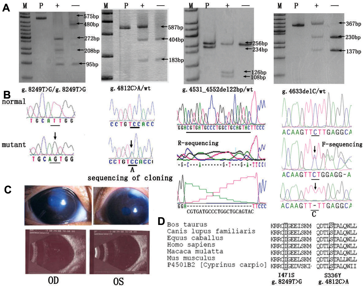
 Figure 1 of
Yang, Mol Vis 2009; 15:432-437.
Figure 1 of
Yang, Mol Vis 2009; 15:432-437.