Figure 7. Immunohistochemical localization of ATM in the adult mouse neuroretina. Immunostaining using a polyclonal antibody raised
against ATM in neuroretina. No specific signal was detected when the specific anti-ATM primary antibody was omitted (A). ATM immunostaining was brown, and sections were counterstained with a methyl green solution. The cellular distribution
of ATM immunostaining was observed in retinal cells (B). A high magnification of the outer part (C) and inner part (D) of the neuroretina is shown. A high magnification of the ganglion cell layer (GCL; E), inner nuclear layer (INL; F), outer nuclear layer (ONL; G) and retinal pigment epithelium (RPE; H) is shown. Abbreviations: choroid (Ch); inner plexiform layer (IPL), outer plexiform layer (OPL), and scleral cells (Sc).
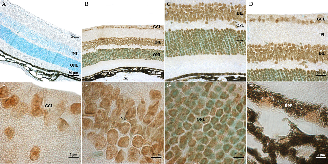
 Figure 7 of
Leemput, Mol Vis 2009; 15:393-416.
Figure 7 of
Leemput, Mol Vis 2009; 15:393-416.