Figure 14. Intracellular localization of ATM and ATMp immunoreactivities in cerebellar Purkinje and granule cells of adult mice using
confocal imaging. We used the monoclonal antibody pS1987 from Rockland for this figure. The same pattern was observed with
rabbit polyclonal antibody pS1987 from Abcam and rabbit polyclonal pS1987 antibody from Signalway. The double labeling of
cerebellar Purkinje cells with ATM immunostaining (green) and PI staining (red; A) is shown. The colocalization in cerebellar granule cells of ATM immunostaining (green) and PI staining (red) is shown. Two-dimensional
scatterplots of voxel intensities in red and green channels are shown in the right-hand column. The Pearson coefficient is
0.42. PI staining and ATM immunoreactivity were colocalized in cerebellar granule cells (B). The colocalization of ATMp immunostaining (green) and PI staining (red) in cerebellar granule cells is also shown. Two-dimensional
scatterplots of voxel intensities in red and green channels are shown in the right-hand column. The Pearson coefficient is
−0.03. PI staining and ATMp antibody staining were not colocalized (C). Abbreviations: Granular cell layer (GL), and Purkinje cell layer (PL).
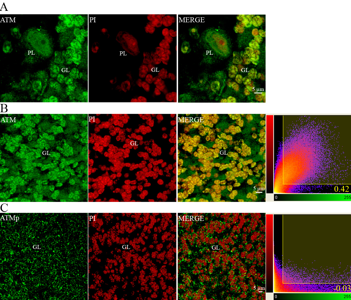
 Figure 14 of
Leemput, Mol Vis 2009; 15:393-416.
Figure 14 of
Leemput, Mol Vis 2009; 15:393-416.