Figure 13. Intracellular localization of ATM and ATMp immunoreactivities in photoreceptors of adult mice using confocal imaging. The
double staining of the ONL using a specific polyclonal antibody raised against ATM (green) in the neuroretina and TO-PRO-3
counterstaining (blue) is shown (A). The double immunostaining with anti-ATM (green) and anti-recoverin (red) antibodies is also shown. Recoverin is a specific
cytoplasmic photoreceptor protein. Two-dimensional scatterplots of voxel intensities in red and green channels are shown in
the right-hand column. We used a threshold of 30 (on a scale 0–255) for each label. Pearson’s correlation coefficient was
determined for the correlation of voxel intensity between the red and green channels and is displayed in the lower right-hand
corner. The Pearson coefficient is −0.28. No colocalization was observed between ATM and recoverin immunostainings in photoreceptor
cells (B). The double immunostaining using an antibody raised against ATMp (green) and recoverin (red) in ONL is clearly visualized.
Two-dimensional scatterplots of voxel intensities in red and green channels are shown in the right-hand column. The Pearson
coefficient is 0.90. ATMp immunostaining colocalized extensively with recoverin immunostaining in photoreceptor cells (C). Abbreviations: Inner nuclear layer (INL), and outer nuclear layer (ONL).
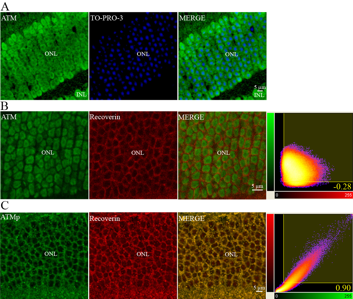
 Figure 13 of
Leemput, Mol Vis 2009; 15:393-416.
Figure 13 of
Leemput, Mol Vis 2009; 15:393-416.