Figure 12. Immunohistochemical localization of ATMp in the adult mouse cerebellar tissue sections. We again used the rabbit polyclonal
pS1987 antibody from Signalway for this figure. The same pattern was observed with the rabbit polyclonal antibody pS1987 from
Abcam and mouse monoclonal antibody pS1987 from Rockland. No specific signal was detected in control experiments where the
specific anti-ATMp antibody was omitted (A). ATMp immunolabeling was detected in specific adult mouse cerebellum neuronal layers (B). A high magnification of the granular cell (GL), molecular cell (ML), and Purkinje cell (PL) layers (C-D) is shown. The ATMp immunostaining of some Pukinje cells axons is clearly visible. A higher magnification of the Purkinje
cell layer is also shown (PL; E).
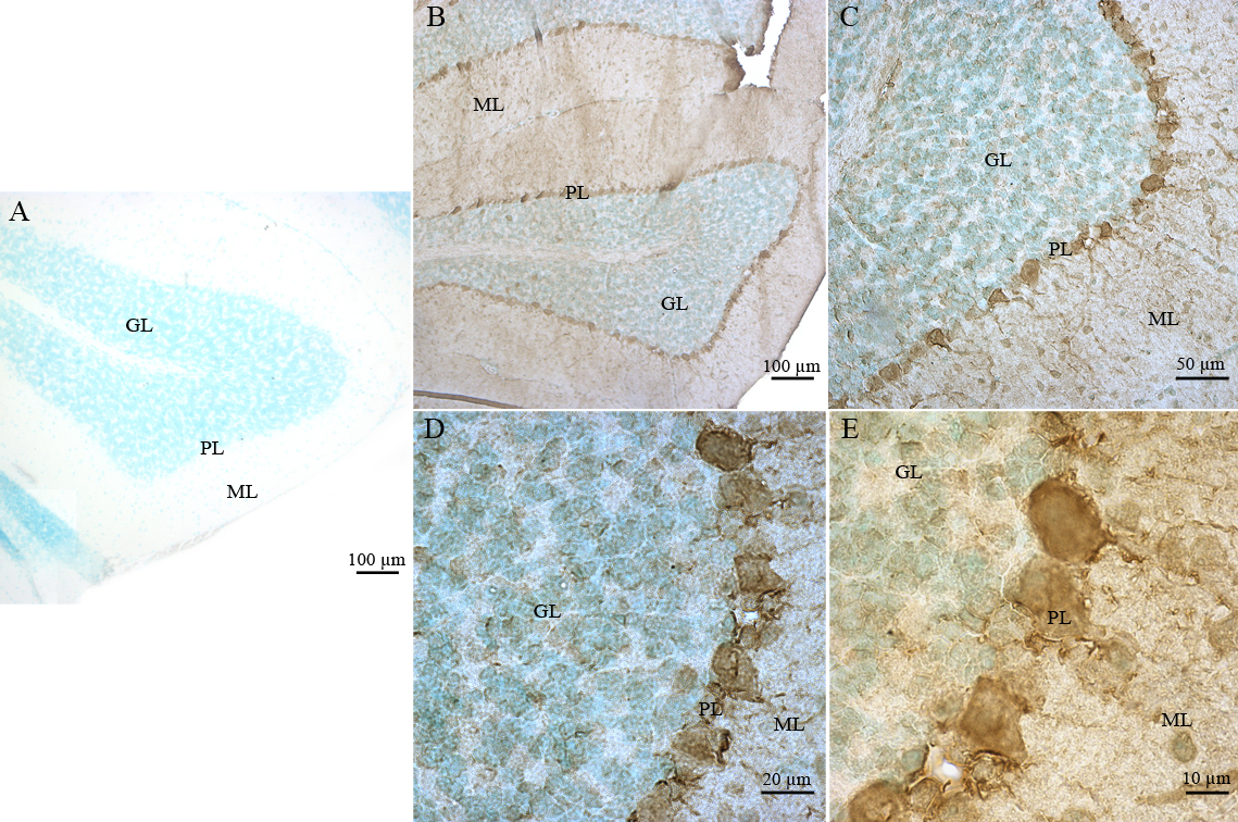
 Figure 12 of
Leemput, Mol Vis 2009; 15:393-416.
Figure 12 of
Leemput, Mol Vis 2009; 15:393-416.