Figure 10. Immunohistochemical localization of ATMp in neuroretinal cells of adult mouse tissue sections. Specific mouse neuroretinal
ATMp immunostaining was detected in adult mouse ocular tissue sections, using an antibody raised against ATMp (the rabbit
polyclonal pS1987 antibody from Signalway was used for this figure). The same pattern was observed with rabbit polyclonal
antibody pS1987 from Abcam and mouse monoclonal antibody pS1987 from Rockland. No specific immunoreactivity was detected in
control experiments where the specific primary antibody was omitted (A). ATMp immunostaining appears with a brown color and sections were counterstained with a methyl green solution. Cellular
distribution of ATMp immunostaining was observed in retinal cells (B). A high magnification of the outer part (C) and inner part (D) of the neuroretina is shown. A high magnification of the ganglion cell layer (GCL; E), inner nuclear layer (INL; F), outer nuclear layer (ONL; G), and retinal pigment epithelium (RPE; H) is also shown. Abbreviations: choroid cells (Ch), inner plexiform layer (IPL), outer plexiform layer (OPL), photoreceptor
inner segment (PIS), and scleral cells (Sc).
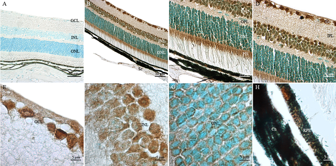
 Figure 10 of
Leemput, Mol Vis 2009; 15:393-416.
Figure 10 of
Leemput, Mol Vis 2009; 15:393-416.