Figure 1. Immunohistochemical localization of 8-oxoguanine and γH2AX. Specific mouse 8-oxoguanine (8-oxo-dG) immunostaining was detected
in adult mouse ocular and cerebellar tissue sections. The cellular distribution of 8-oxo-dG immunostaining was observed in
retinal cells of C57BL/6J (A), Atm+/+ (B), and Atm−/− mice (C). No specific retinal (D) or cerebellar (E) signal could be detected in control experiments. The 8-oxo-dG immunostaining was also observed in cerebellar cells (F). A high magnification of 8-oxo-dG immunostaining in the Purkinje cell layer (PL; G) is shown. Specific mouse γH2AX immunostaining was detected in adult mouse ocular tissue sections. No specific immunoreactivity
was detected in control experiments (H). γH2AX immunostaining was observed in C57BL/6J (I) Atm+/+ (J) and Atm−/− mouse retina (K). Abbreviations: ganglion cell layer (GCL); granular cell layer (GL), inner nuclear layer (INL); molecular cell layer (ML),
outer nuclear layer (ONL), and photoreceptor inner segment (PIS).
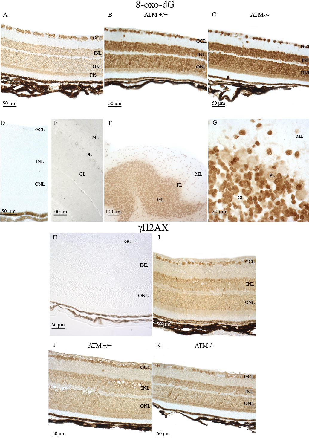
 Figure 1 of
Leemput, Mol Vis 2009; 15:393-416.
Figure 1 of
Leemput, Mol Vis 2009; 15:393-416.