Figure 3. Forward and reverse sequence analysis of the affected and unaffected individuals in this ADCC Chinese family. It shows a heterozygous
mutation (IVS3–1 G>A) in the third canonical AG sites of MIP (black triangles). The black vertical line denotes the normal intron 3-exon 4 acceptor splice site. The mutation IVS3 −1
G>A abolishes a BstSF I site (underlined) which is enzymatic cut indicated by the arrow.
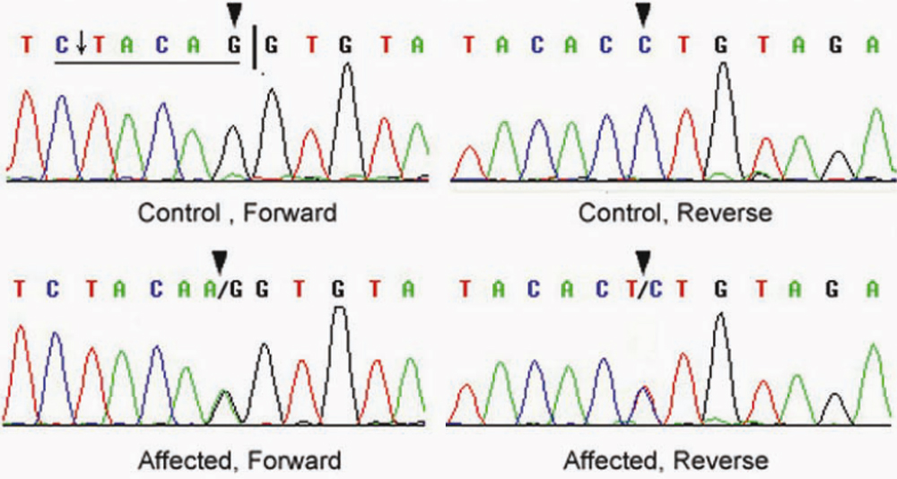
 Figure 3 of
Jiang, Mol Vis 2009; 15:38-44.
Figure 3 of
Jiang, Mol Vis 2009; 15:38-44.