Figure 2. Photographs of the left eye of the proband with congenital cataract. A: Diffuse illumination shows a “snail-like” cataract with opacity density gradually increased from the peripheral adult nucleus
to the inner embryonal nucleus, while the cortex remains transparent. B: Slit section shows that the opacified nuclei are separated by a transparent circle (white arrow).
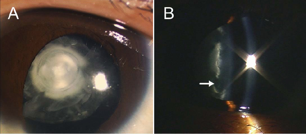
 Figure 2 of
Jiang, Mol Vis 2009; 15:38-44.
Figure 2 of
Jiang, Mol Vis 2009; 15:38-44.