Figure 3. Light microscope images of normal and injured bovine corneas. Cross-sectional images taken from the center of a normal/uninjured
bovine cornea (A) and a bovine cornea immediately after injury (0 h) and at 1 week post injury (B). Black arrows represent the wound edge. Immediately after injury stromal gaps are evident at the wound edge. After 1 week
of wound healing the stromal gaps are smaller and epithelial thickening is evident in the wound area. Scale bars for images
A and B are 100 μm and 25 μm, respectively.
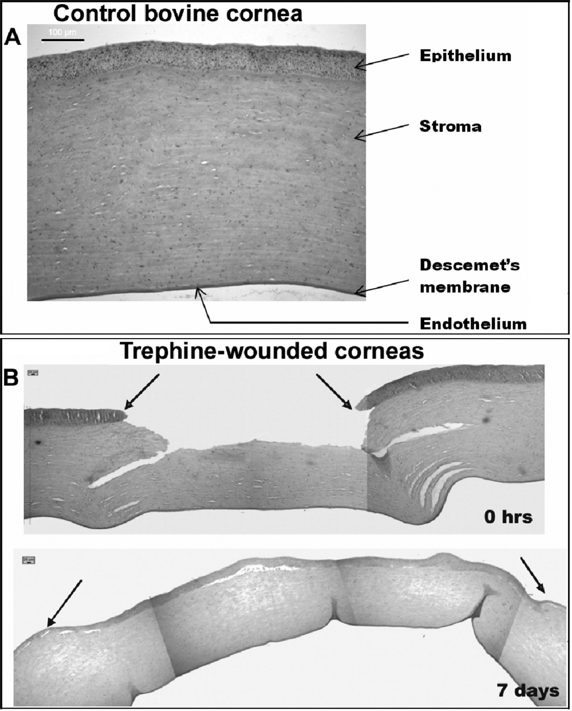
 Figure 3 of
Kamma-Lorger, Mol Vis 2009; 15:378-385.
Figure 3 of
Kamma-Lorger, Mol Vis 2009; 15:378-385.