Figure 1 of
Kamma-Lorger, Mol Vis 2009; 15:378-385.
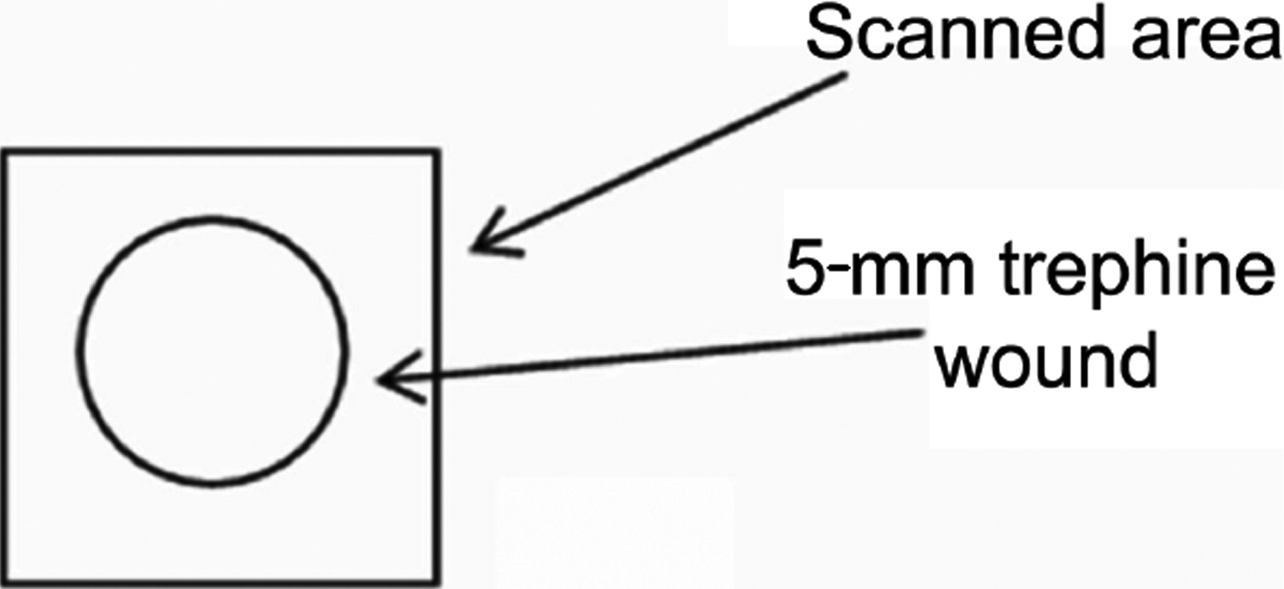
Figure 1.
Square corneal piece from central cornea with the 5 mm trephine wound in the middle. The whole area of the corneal piece was scanned.
 Figure 1 of Kamma-Lorger, Mol Vis 2009; 15:378-385. Figure 1 of Kamma-Lorger, Mol Vis 2009; 15:378-385.