Figure 6. Analysis of GPR109A mRNA and
protein expression in primary RPE cells isolated from mouse retina. A:
This phase-contrast image is representative of the morphology of mouse
primary retinal pigment epithelium (mRPE) after 6 days in culture. B:
In this panel, the morphology of mRPE cells is shown at higher
magnification. C: mRPE cells incubated with antibody against
CRALBP were positive for this RPE/Müller cell marker. D: mRPE
cells incubated with antibody against the RPE-specific cellular marker,
RPE-65, were highly positive. E: RT–PCR analysis of RNA
collected from mRPE cells detected robust GPR109A mRNA expression. F:
Incubation of mRPE cells with anti-GPR109A antibody yielded positive
signals. G: No signal was detected in mRPE cells incubated with
primary GPR109A antibody neutralized by incubation with excess
antigenic peptide.
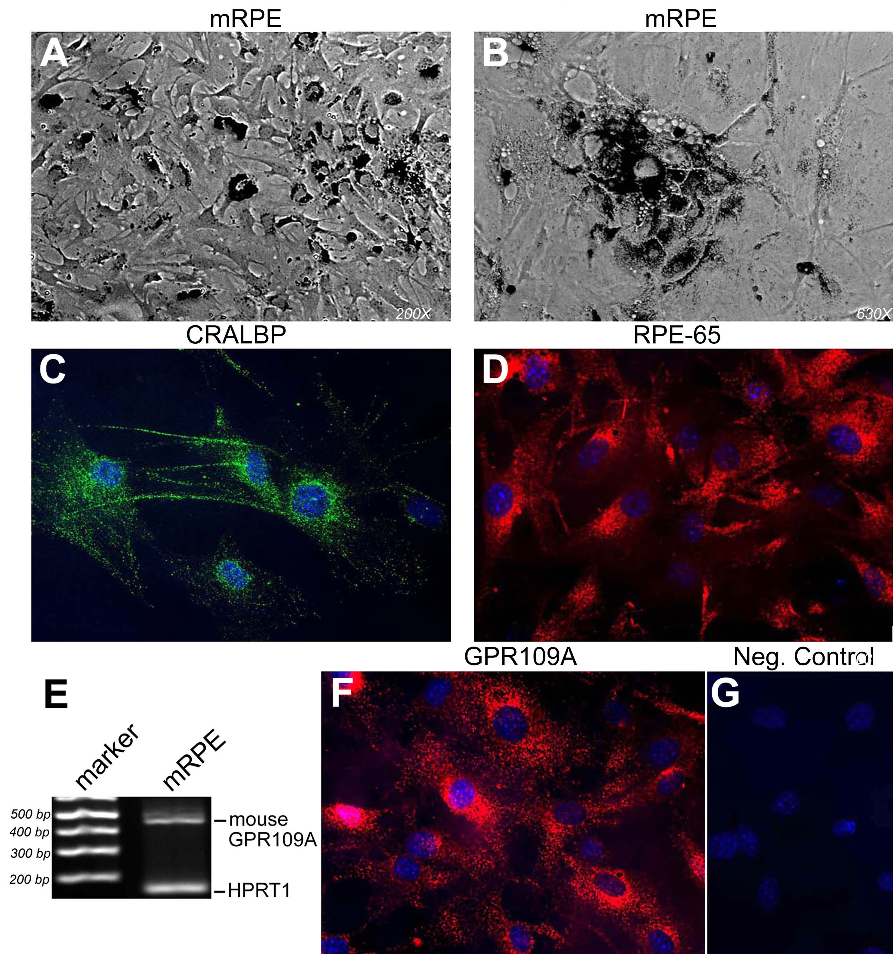
 Figure 6 of Martin, Mol Vis 2009; 15:362-372.