Figure 5. Immunofluorescence analysis of
GPR109A protein in ARPE-19, rMC-1, and RGC-5 cells. A: ARPE-19
cells incubated with anti-GPR109A antibody, which detects both GPR109A
and GPR109B, were highly positive. B: Cells incubated with
primary GPR109A antibody neutralized by incubation with excess
antigenic peptide did not display any positive signal. Likewise, no
positive signal was detected in rat Müller (rMC-1; C) and
retinal ganglion-5 (RGC-5; D) cells incubated with anti-
GPR109A antibody.
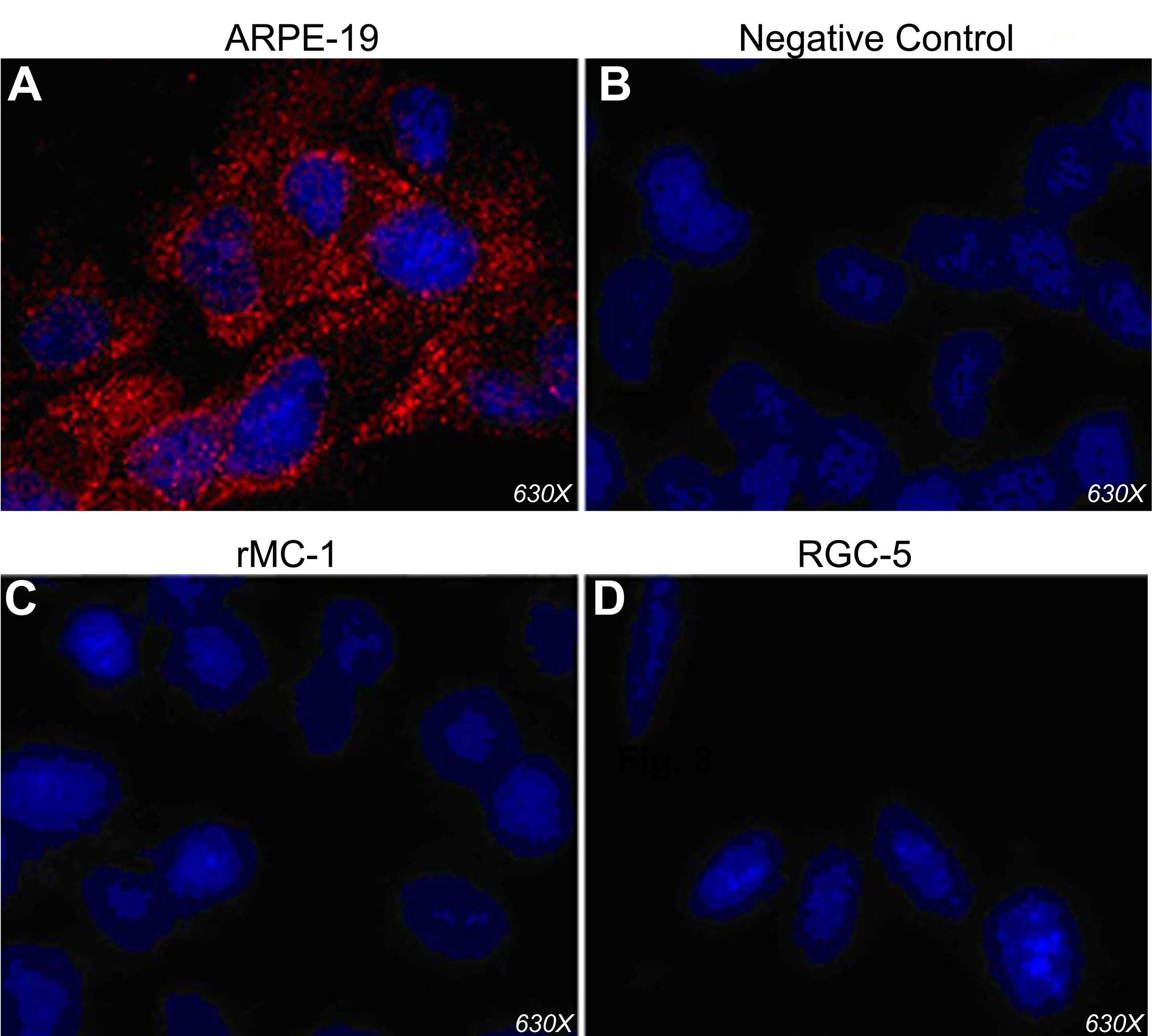
 Figure 5 of Martin, Mol Vis 2009; 15:362-372.