Figure 3. Double-labeling
immunofluorescence techniques were employed to determine whether
GPR109A protein is expressed apically or basolaterally in RPE. Chicken
polyclonal anti-monocarboxylate transporter 1 (MCT1) was used as a
positive marker for RPE apical membrane. Sections were then viewed and
optically sectioned using a Zeiss Axioplan fluorescent microscope
equipped with an apotome. Merging of positive signals for MCT1 (red)
and GPR109A protein (green) did not display any significant overlap.
Hoechst 33342 nuclear stain is shown in blue.
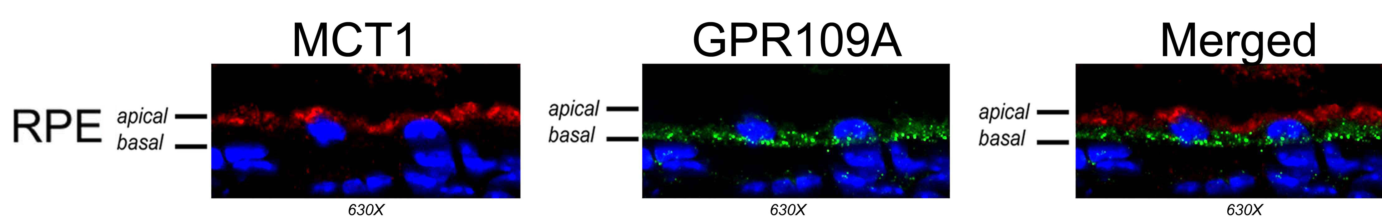
 Figure 3 of Martin, Mol Vis 2009; 15:362-372.