Figure 2. Localization of GPR109A protein
in intact retina. A: A hematoxylin and eosin-stained retinal
cryosections is provided for comparison to panel B of the
figure. B: Incubation of mouse retinal cryosections with anti-
GPR109A antibody revealed positive (red) labeling for GPR109A protein
exclusively in retinal pigment epithelium (RPE). Higher magnification
of cryosections reveals that the GPR109A protein is associated
specifically with RPE basolateral membrane (panel B, inset).
Hoechst 33342 nuclear stain is shown in blue. In the inset, Nu means
nucleus. C: No labeling was detected in retinal cryosections
incubated with primary GPR109A antibody neutralized by incubation with
excess antigenic peptide.
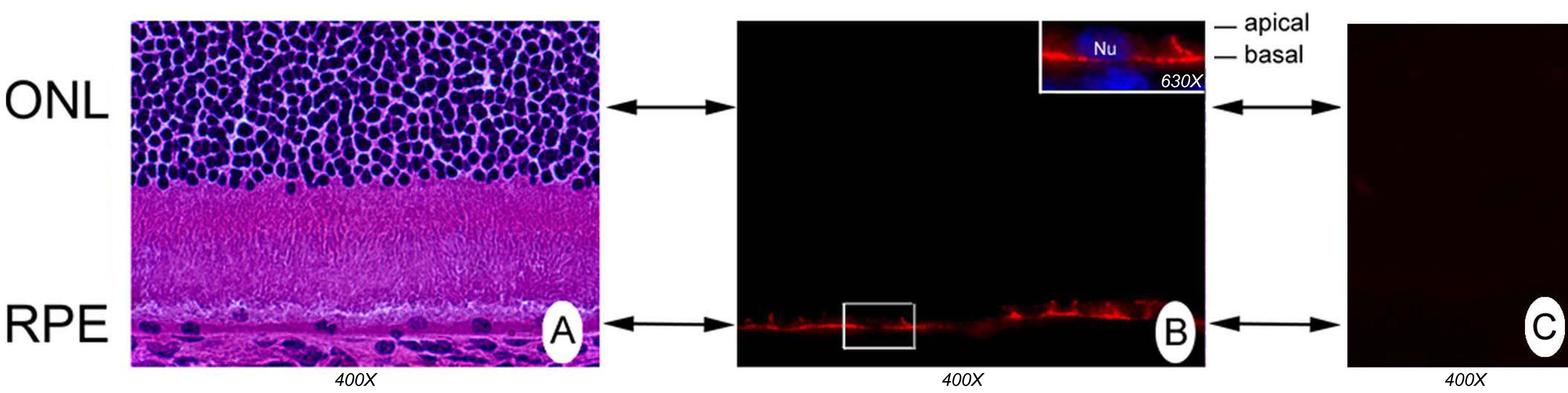
 Figure 2 of Martin, Mol Vis 2009; 15:362-372.