Figure 8. Quantification of TUNEL-positive
cells in the outer nuclear layer of affected dogs. Apoptosis analysis
was performed with fluorescent microscopy and a 40× objective. Terminal
dUTP nick end labeling (TUNEL) positive cells stained with fluorescein
were visualized and counted. TUNEL-positive cells in the outer nuclear
layer (ONL) were quantified as the percent of the total number of cells
in ONL per field. Data are the mean±standard error of the mean from 10
fields of view. The graph represents the average of 3 independent
experiments. Months are abbreviated as m.
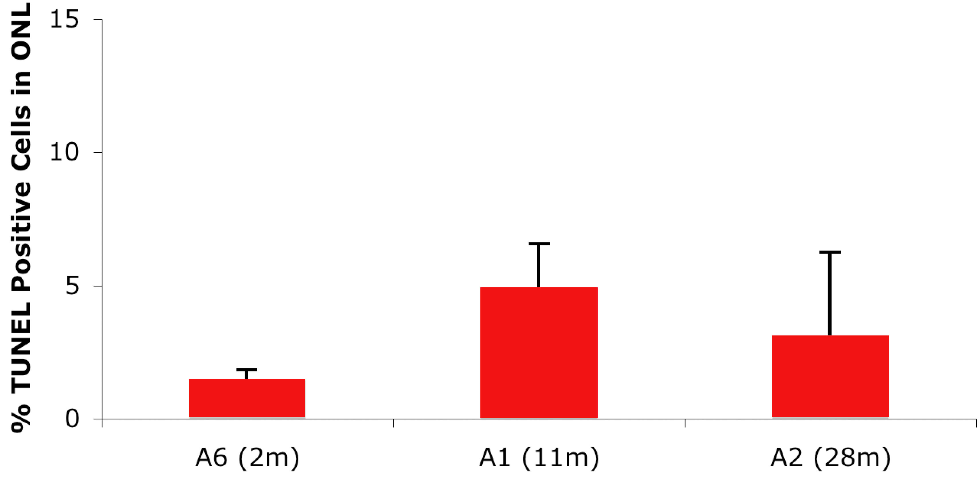
 Figure 8 of Lhériteau, Mol Vis 2009; 15:349-361.