Figure 7. TUNEL assay on normal and
affected dogs. A: Retina of the normal dog NA2 displayed no
TUNEL-positive labeling. B: Retina of the affected dog A6 at
the age of 2 months treated with DnaseI (positive control). C:
Retina of the affected dog A6 at the age of 2 months treated with label
solution only (negative control). TUNEL staining (D,E,F) and
corresponding light micrographs (G,H,I) of the retinas of
affected dogs. (D,G) retina of the affected dog A6 at the age of
2 months, (E,H) retina of the affected dog A1 at the age of 11
months and (F,I) retina of the affected dog A2 at the age of 28
months. TUNEL staining revealed apoptotic cells in all affected dogs,
mostly in the outer nuclear layer, indicating apoptosis of
photoreceptor cells. Abbreviations: retinal pigment epithelium (RPE);
outer segments (OS); inner segments (IS); outer nuclear layer (ONL);
outer plexiform layer (OPL); inner nuclear layer (INL); inner plexiform
layer (IPL); ganglion cell layer (GCL). The scale bar represents 100 µm.
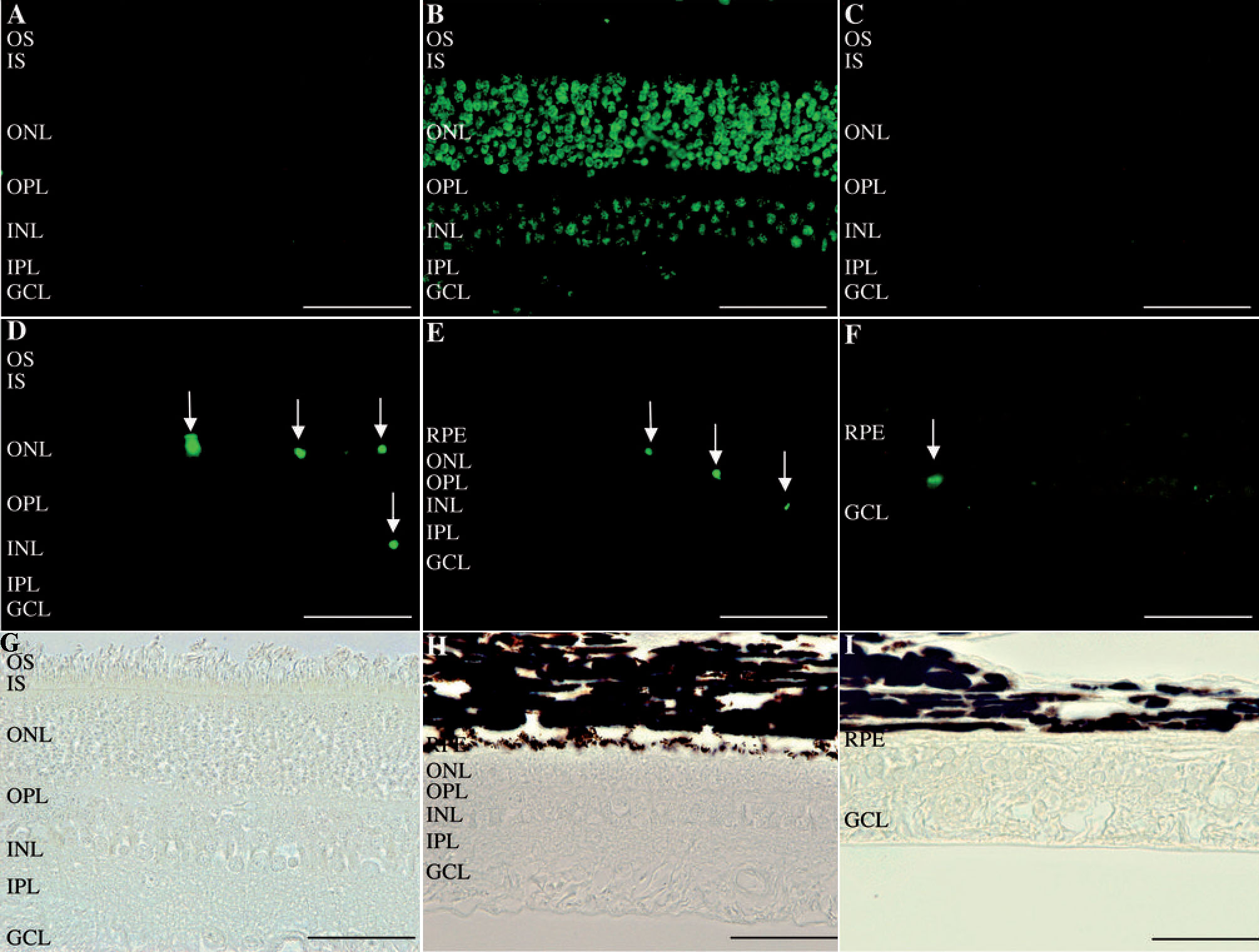
 Figure 7 of Lhériteau, Mol Vis 2009; 15:349-361.