Figure 6. Light micrographs and
immunofluorescent staining. A,B,C,D: Retina of the non affected
dog NA2. E,F,G,H: Retina of the 2 months old affected dog A6. B,F:
Rod labeling with anti-Rho4D2 antibody. D,H: Cone labeling with
PNA-FITC lectin. These results show that both the rods and cones are
still present at the age of 2 months in affected dogs. Abbreviations:
retinal pigment epithelium (RPE); outer segments (OS); inner segments
(IS); outer nuclear layer (ONL); outer plexiform layer (OPL); inner
nuclear layer (INL); inner plexiform layer (IPL); ganglion cell layer
(GCL). The scale bar represents 50 µm.
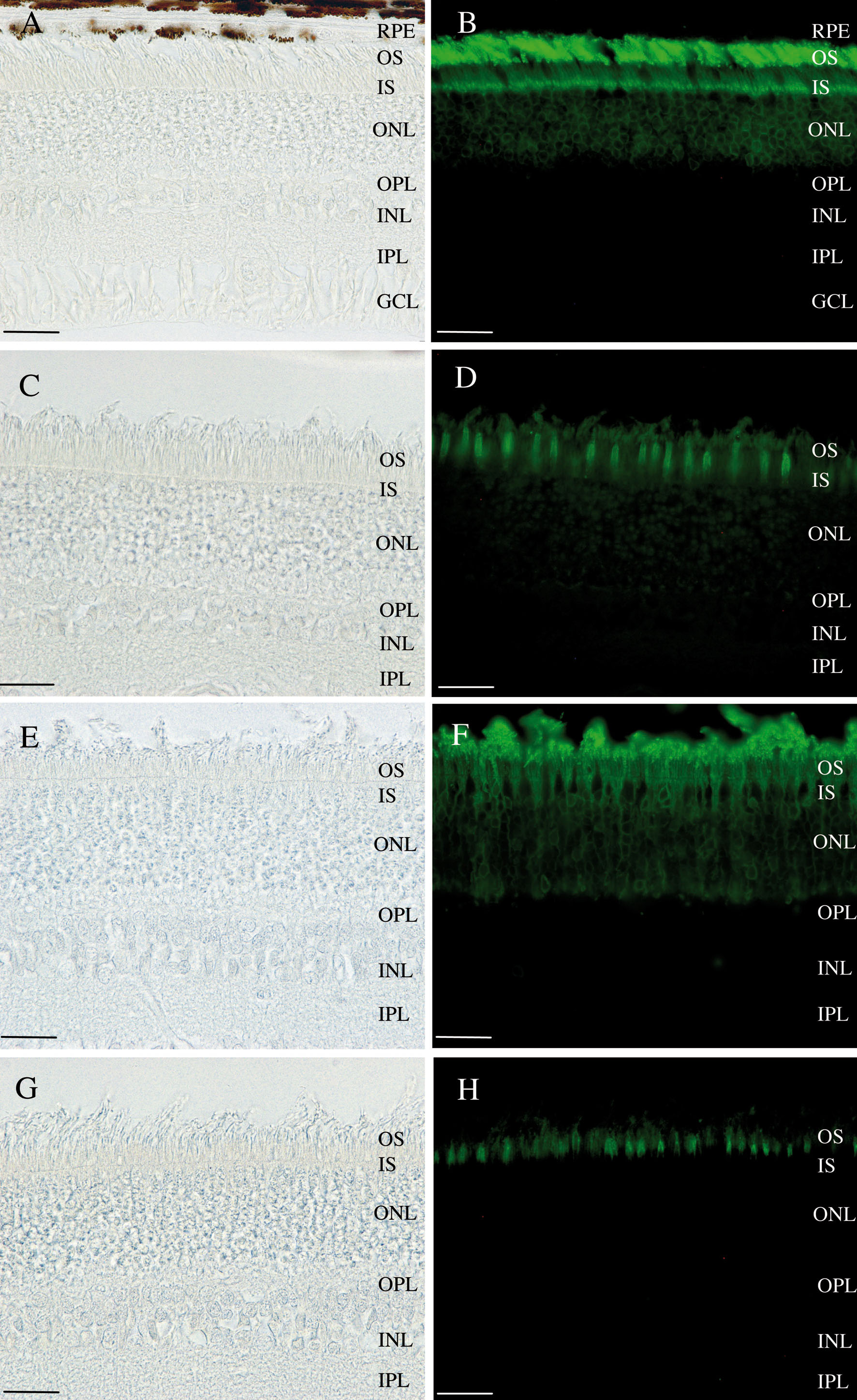
 Figure 6 of Lhériteau, Mol Vis 2009; 15:349-361.