Figure 5. Histological sections of RPGRIP1-deficient
dog’s retinas. A: Normal dog NA2 at the age of 2 months. B:
Affected dog A6 at the age of 2 months. C: Affected dog A1 at
the age of 11 months. D: Affected dog A2 at the age of 28
months. E: Affected dog A5 at the age of 10 years. These
results confirm the thinning of the retina observed on OCT images.
Abbreviations: retinal pigment epithelium (RPE); outer segments (OS);
inner segments (IS); outer nuclear layer (ONL); outer plexiform layer
(OPL); inner nuclear layer (INL); inner plexiform layer (IPL); ganglion
cell layer (GCL). The scale bar represents 100 µm.
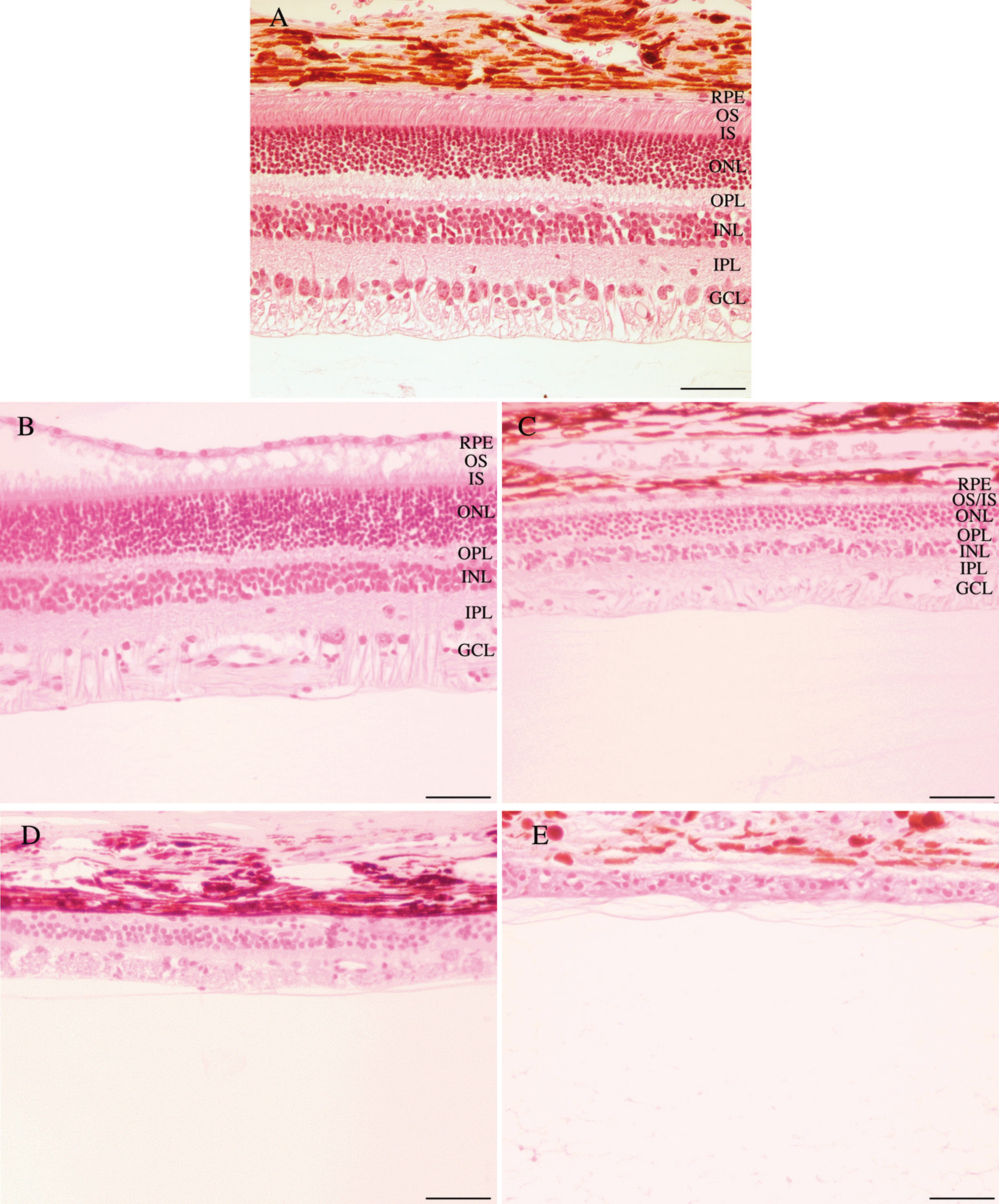
 Figure 5 of Lhériteau, Mol Vis 2009; 15:349-361.