Figure 3. Images of OCT recordings of RPGRIP1-deficient dogs. The insets show the localization of the 3mm horizontal line scan above the optical nerve head. Notable decrease
of the retinal thickness is observed in the affected dog A2 between 5 (A) and 12 months (B). Surprisingly, extreme thinning is seen in the 10-year-old affected dog A5 (C).
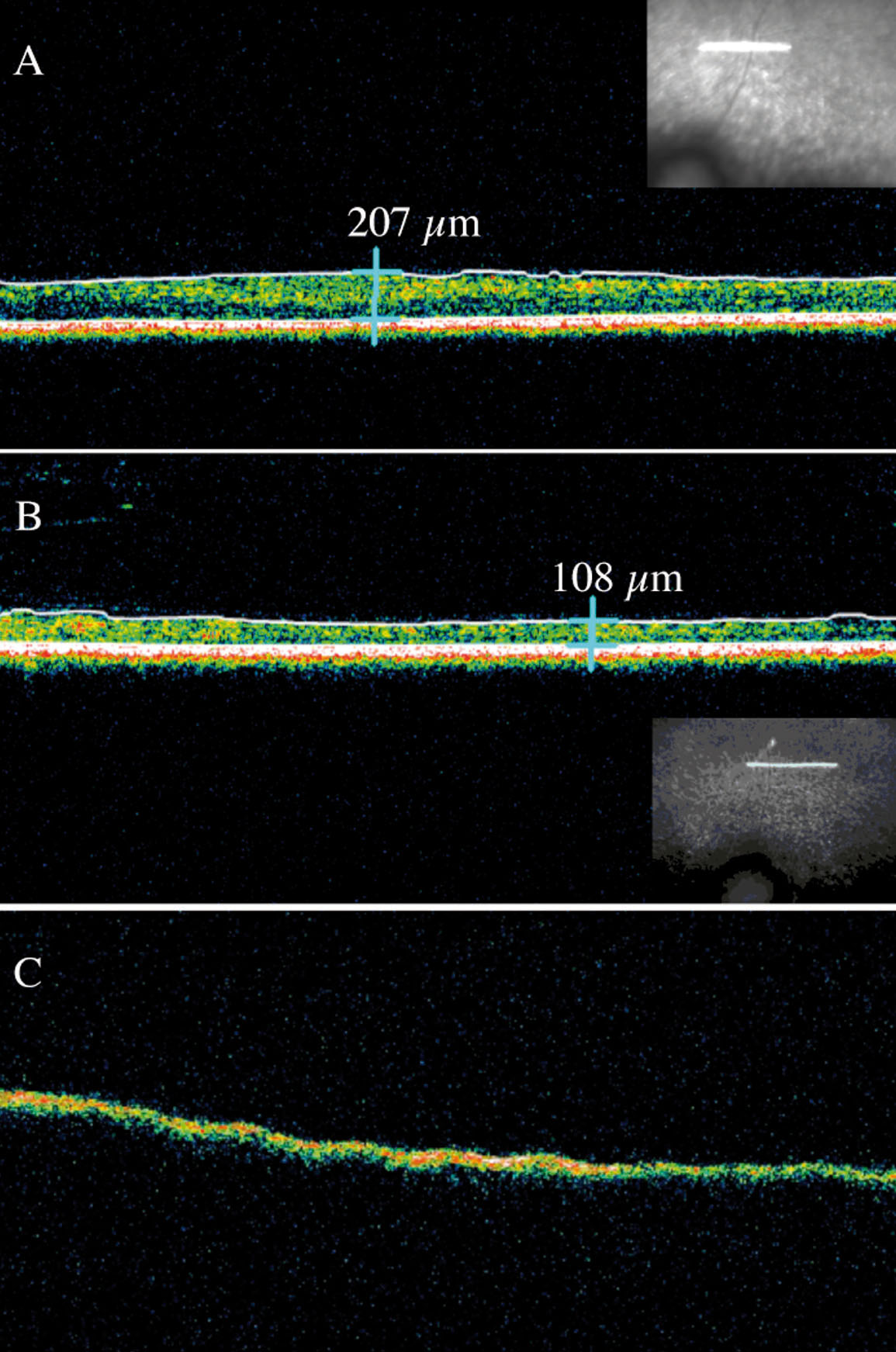
 Figure 3 of
Lhériteau, Mol Vis 2009; 15:349-361.
Figure 3 of
Lhériteau, Mol Vis 2009; 15:349-361.