Figure 2. Western blot analysis of S100 proteins expressed in human conjunctiva and pterygium tissue. Uninvolved conjunctiva (Lane 1)
and pterygium (Lane 2) tissues were lysed in RIPA buffer and 60 μg of total protein was loaded in each lane of SDS-PAGE and
transferred to nitrocellulose membrane and probed with antibodies against various S100 proteins as well as β-actin (loading
control). Bands of 11 kDa corresponding to S100A4, S100A6, and S100A11 were detected. Bands corresponding to the molecular
weight of 8 kDa and 13 kDa confirmed the presence of S100A8 and S100A9, respectively.
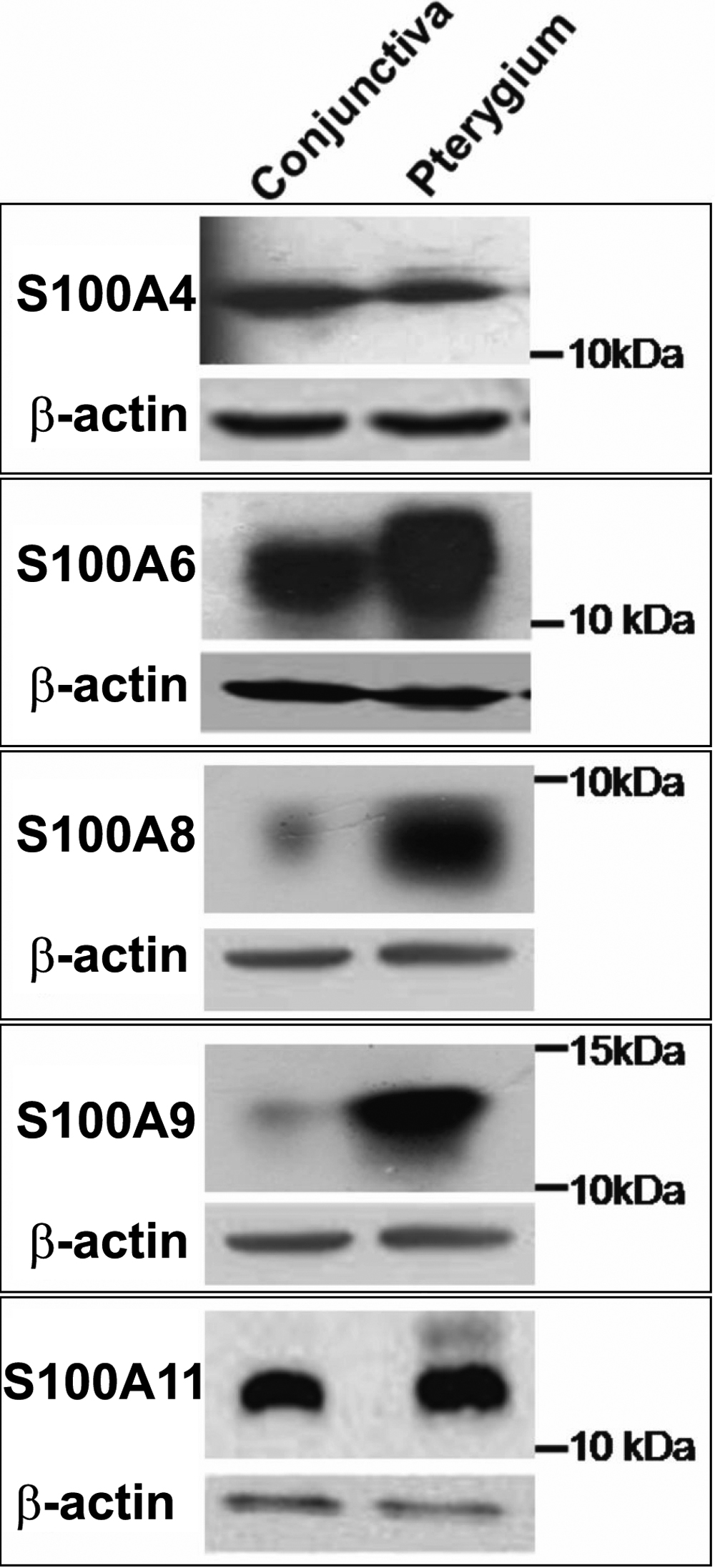
 Figure 2 of
Riau, Mol Vis 2009; 15:335-342.
Figure 2 of
Riau, Mol Vis 2009; 15:335-342.