Figure 1. Immunofluorescent staining of
S100 proteins in human conjunctival and pterygial epithelia. Primary
antibodies against S100A4 (C and D), S100A6 (E
and F), S100A8 (G and H), S100A9 (I and J),
and S100A11 (K and L) were used. Negative controls were
shown in A and B. Images of uninvolved conjunctiva (A,
C, E, G, I, and K) and pterygium
epithelium (B, D, F, H, J, and L)
were shown. The nuclei were stained with DAPI present in the mounting
medium. A dashed line indicates the location of the basement membrane.
All images were taken at 400X magnification.
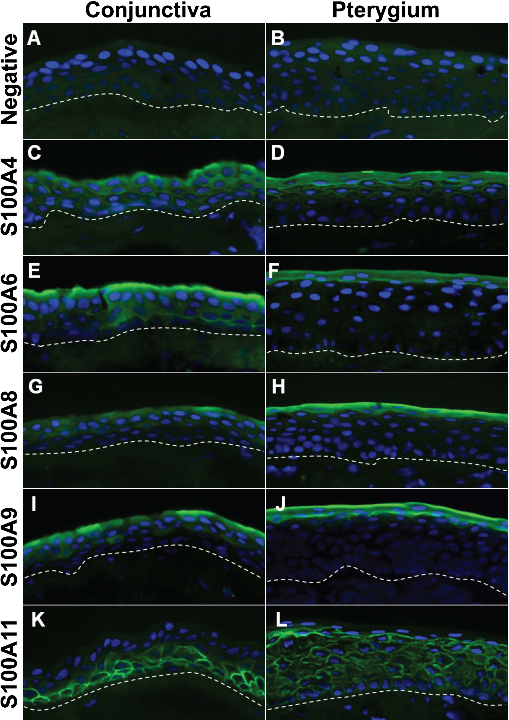
 Figure 1 of Riau, Mol Vis 2009; 15:335-342.