Figure 7. Color fundus photographs and
spectral domain optical coherence tomography scans of patient CT13 and
patient CT12. A normal fovea and an atrophic macular lesion are shown
on color fundus photographs of patient CT13 (A) and patient CT04
(B), respectively. Spectral domain high-definition optical
coherence tomography scan shows, in the macular area of patient CT13, a
thickening of the layer corresponding to the junction between the
retinal pigment epithelium (RPE) and the interface of the inner segment
and outer segment of the photoreceptor (C). A thinning of all
the retinal layers with enhancement of reflectivity of RPE, which seems
to spread far behind it (D), appears on the macular scan of
patients CT12.
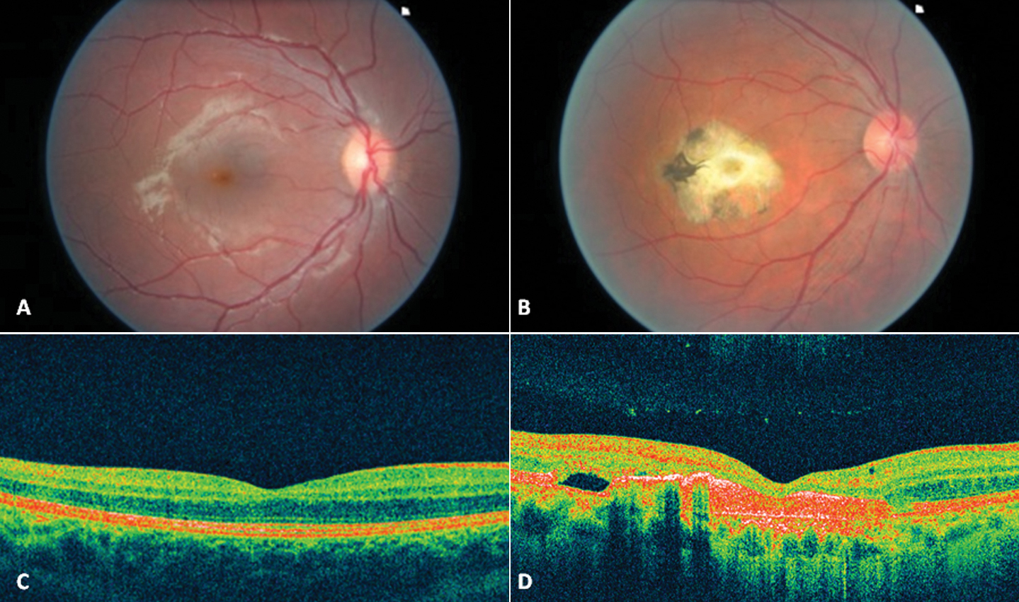
 Figure 7 of Querques, Mol Vis 2009; 15:2960-2972.