Figure 6. Color fundus photographs and
spectral domain optical coherence tomography scans of patient CT02 and
patient CT04. A normal fovea and a vitelliruptive macular lesion are
shown on color fundus photographs of patient CT02 (A) and
patient CT04 (B), respectively. Spectral domain high-definition
optical coherence tomography scan shows, in the macular area of patient
CT02, a thickening of the layer corresponding to the junction between
the retinal pigment epithelium (RPE) and the interface of the inner
segment and outer segment of the photoreceptor (C). An optically
empty lesion between the RPE and the inner segment /outer segment
interface, with clumping of hyper-reflective material on the posterior
retinal surface and, on some parts, a hyper-reflective mottling stuck
on the RPE layer (D), appears on the macular scan of patient
CT04.
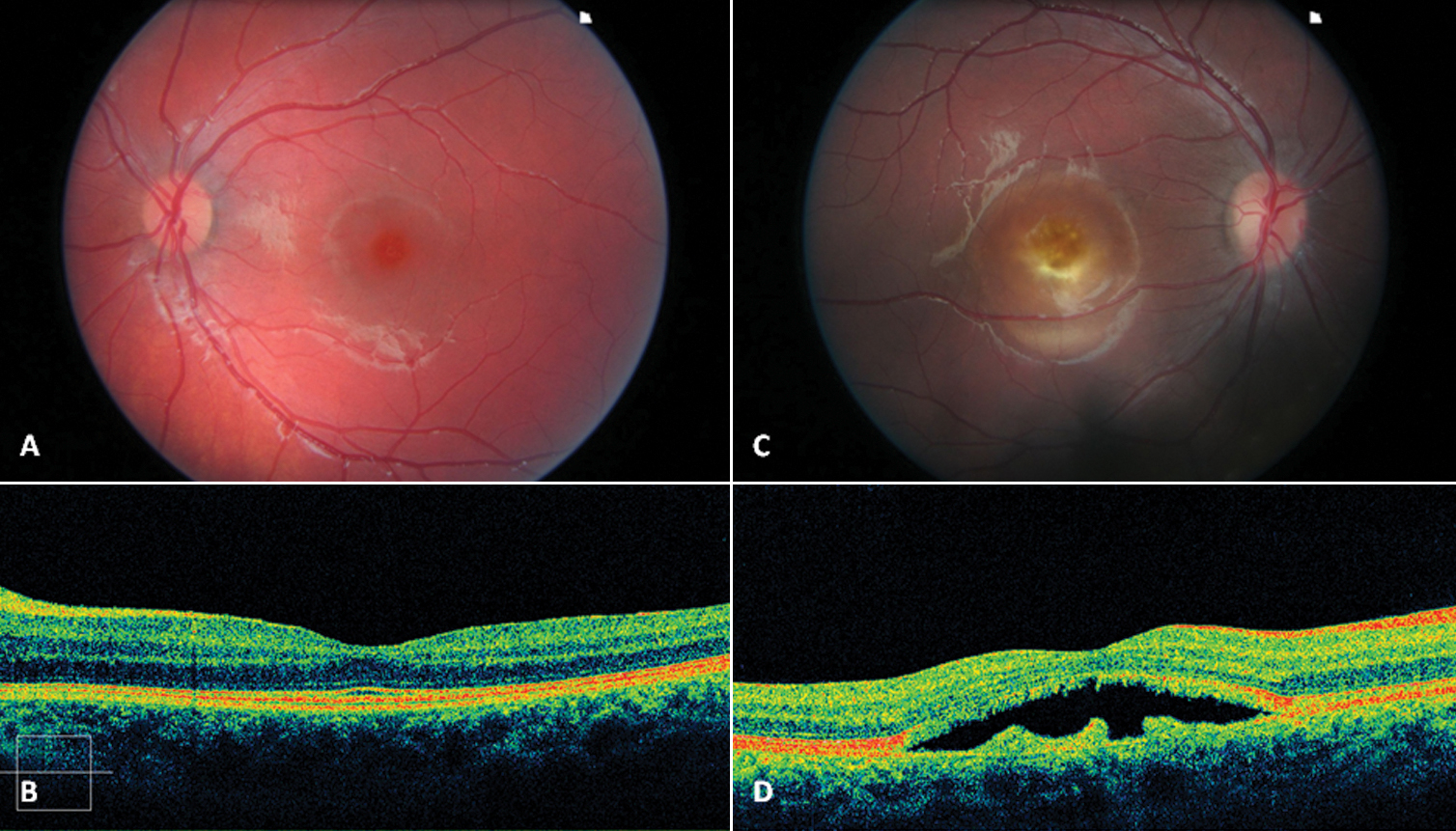
 Figure 6 of Querques, Mol Vis 2009; 15:2960-2972.