Figure 2. Electropherograms of the three
novel BEST1 mutations. These electrophoregrams show an
heterozygous peak GA at position 44 responsible for a p.G15D mutation
in family FG IV, an heterozygous peak TC at position 791 responsible
for a p.I230T mutation in family CT II, and an heteozygous peak AG at
position 10 responsible for a p.T4A mutation in family CT IV.
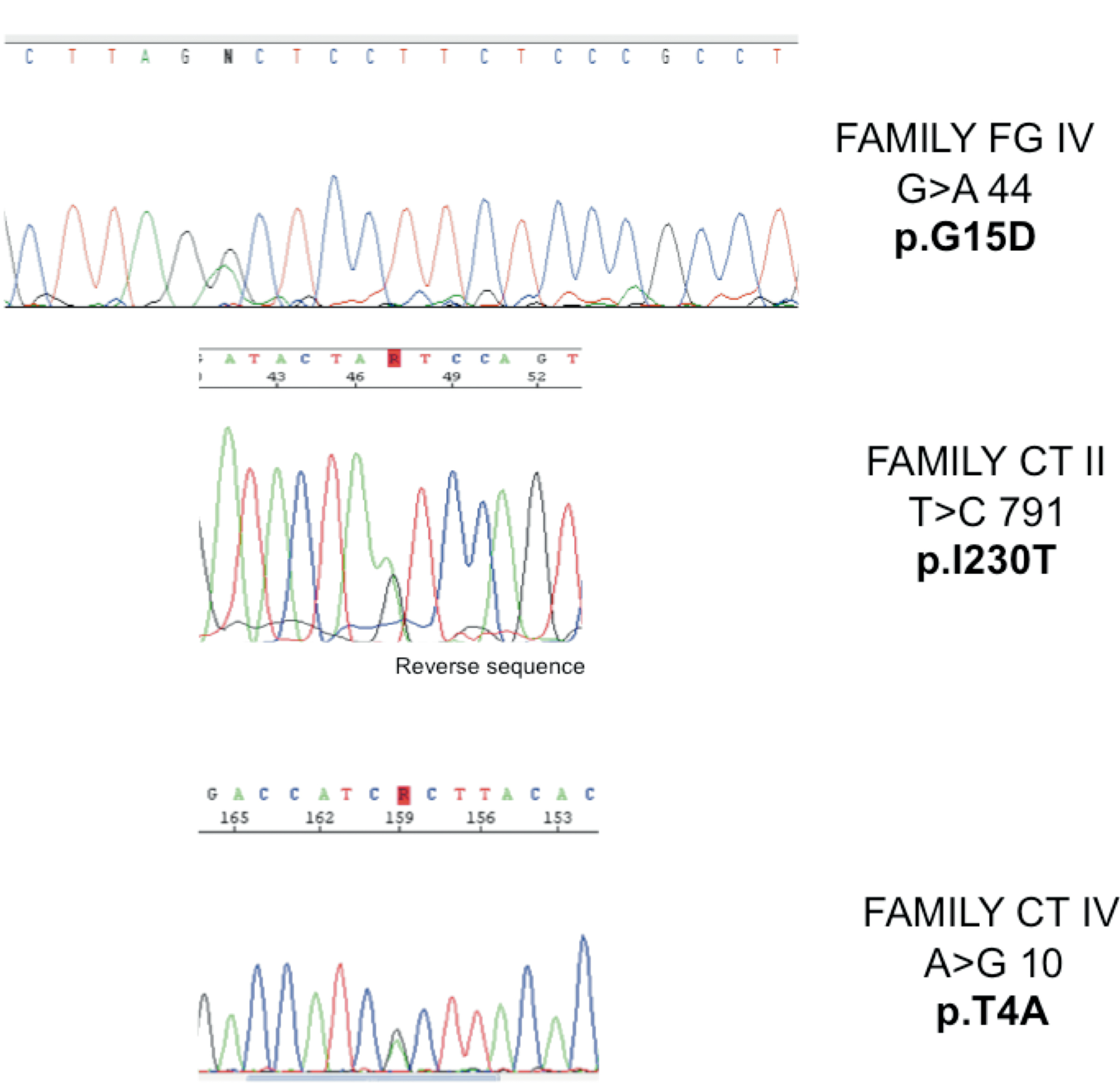
 Figure 2 of Querques, Mol Vis 2009; 15:2960-2972.