Figure 4. The percentage of interleukin-21 receptor positive cells from draining lymph node of experimental autoimmune uveitis and control
mice. The isolated DLN cells were stained with anti-CD4, anti-CD8, and anti-IL-21R Ab for 0.5 h for the analysis of surface
markers. A: Dot plots of IL-21R+ cells in CD4+ and CD8+ cells of EAU and control mice. Data shown are representative of six independent experiments. B: Percentages of IL-21R+ cells in CD4+ T cells of EAU and control mice. C: Percentages of IL-21R+ cells in CD8+ T cells. Data are presented as mean±standard deviation; n=6 per group.
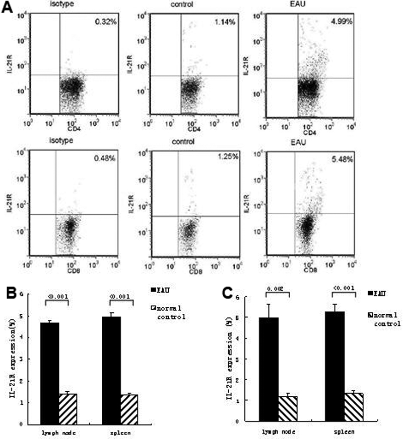
 Figure 4 of
Liu, Mol Vis 2009; 15:2938-2944.
Figure 4 of
Liu, Mol Vis 2009; 15:2938-2944.