Figure 1. Histopathologic features of the eyes of experimental autoimmune uveitis (EAU) mice. Eyes were enucleated from EAU and complete
Freund’s adjuvant (CFA) control mice on day 14 after immunization. Sections (5 µm) were cut through the pupillary–optic nerve
plane and stained with hematoxylin and eosin. A: Eye of CFA controls. The retina layers are well ordered and preserved. B: Eye of EAU mice. The normal structure of the retina is disorganized. Inflammatory cells infiltrate the ciliary body, choroid,
vitreous and retina. Vasculitis, granuloma formation, and retinal folding are visible, and the photoreceptor layer is damaged.
The scale bar=50 µm.
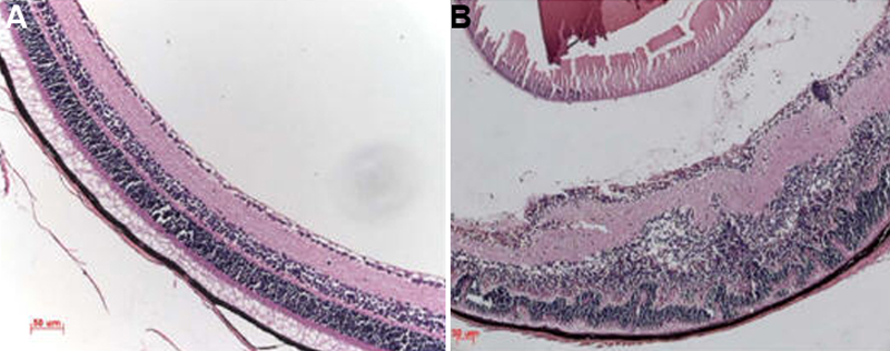
 Figure 1 of
Liu, Mol Vis 2009; 15:2938-2944.
Figure 1 of
Liu, Mol Vis 2009; 15:2938-2944.