Figure 1 of
Zhang, Mol Vis 2009; 15:2911-2918.
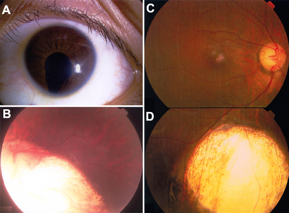
Figure 1.
Uveal coloboma.
A
and
B
demonstrate iris coloboma (
A
) and choroid coloboma involving the optic disc (
B
).
C
and
D
show inferior choroid coloboma. (
D
did not align well with
C
).
 Figure 1 of
Zhang, Mol Vis 2009; 15:2911-2918.
Figure 1 of
Zhang, Mol Vis 2009; 15:2911-2918.