Figure 4. Histopathologic examination of S-1-induced corneal disorders. A: Sections were subjected to hematoxylin and eosin (HE) staining and to immunofluorescence staining for both cytokeratin 12
(K12, in green) and cytokeratin 4 (K4, in red). HE staining revealed that each epithelial sheet lacked the stratified structure
of the normal corneal epithelium. Immunofluorescence analysis revealed the presence of four types of cells in each lesion:
those positive for one, both, or neither of the two cytokeratins. Nuclei were stained with 4′,6-diamidino-2-phenylindole (blue).
B: K12 was only expressed in corneal epithelium, whereas K4, by contrast, was only expressed in conjunctival epithelium in
immunofluorescence staining of the normal human cornea and conjunctiva for K12 and K4.
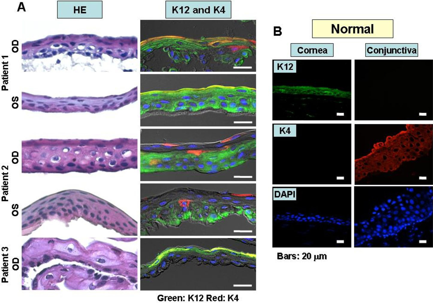
 Figure 4 of
Chikama, Mol Vis 2009; 15:2896-2904.
Figure 4 of
Chikama, Mol Vis 2009; 15:2896-2904.