Figure 3. Inflammatory changes in ocular mucositis revealed by in vivo confocal microscopy. In vivo confocal microscopy revealed a high
density of dendritic and spherical cells, likely including Langerhans cells, macrophages, monocytes, and neutrophils, in the
epithelial basal cell layer (A, B, D) as well as many activated keratocytes in the anterior stroma (C) and a reduced number of subepithelial nerve fibers (F). Fibrosis (arrows) with a circular pattern of degradation of Bowman’s layer was observed in a case that subsequently showed
only minor improvement after discontinuation of S-1 administration (E).
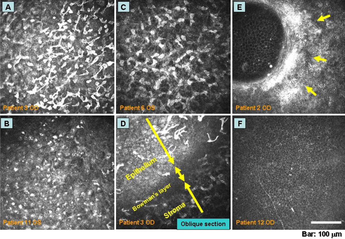
 Figure 3 of
Chikama, Mol Vis 2009; 15:2896-2904.
Figure 3 of
Chikama, Mol Vis 2009; 15:2896-2904.