Figure 1. In vivo confocal microscopy of the cornea and the limbus in a normal eye with the an in vivo laser confocal microscope with
an attachment for the cornea (Heidelberg Retina Tomograph II–Rostock Cornea Module, or HRTII-RCM®; Heidelberg Engineering,
Heidelberg, Germany). Panels A through F represent areas of 400×400 µm corresponding to the superficial epithelial cell layer, wing cell layer, basal cell layer,
Bowman’s layer, mid stroma, and endothelium, respectively. The approximate levels of the sections are indicated in the hematoxylin
and eosin-stained histologic specimen. Panels G and H show the limbus at the border between the cornea and conjunctiva and the limbal palisades of Vogt, respectively. The peripheral
cornea often retains melanocytes in the basal epithelial layer (inset in G).
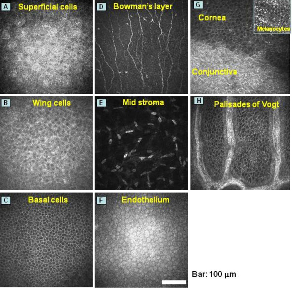
 Figure 1 of
Chikama, Mol Vis 2009; 15:2896-2904.
Figure 1 of
Chikama, Mol Vis 2009; 15:2896-2904.