Figure 3. Nuclear translocation of NFκB
subunits upon viral infection. Confocal microscopy shows cellular
localization of NFκB subunits. A: Cytoplasmic localization of
NFκB p65 (yellow) is compared to nuclei (blue, DAPI stain) in
mock-infected keratocytes (M). Adenoviral-infected (V) keratocytes
demonstrate nuclear localization of NFκB p65 and p50. Nuclear
translocation of NFκB p65 was reduced in the presence of inhibitors of
p38 (SB) and ERK (PD). The nuclear localization of both NFκB p65 and
p50 was partially reduced upon pretreatment with an inhibitor of JNK
(SP) and completely blocked with an inhibitor of Src (PP2). Isotype
control is shown (Ctrl). Viral infection also induced nuclear
translocation of cREL (B), while all signaling inhibitors tested
reduced the nuclear translocation of cREL.
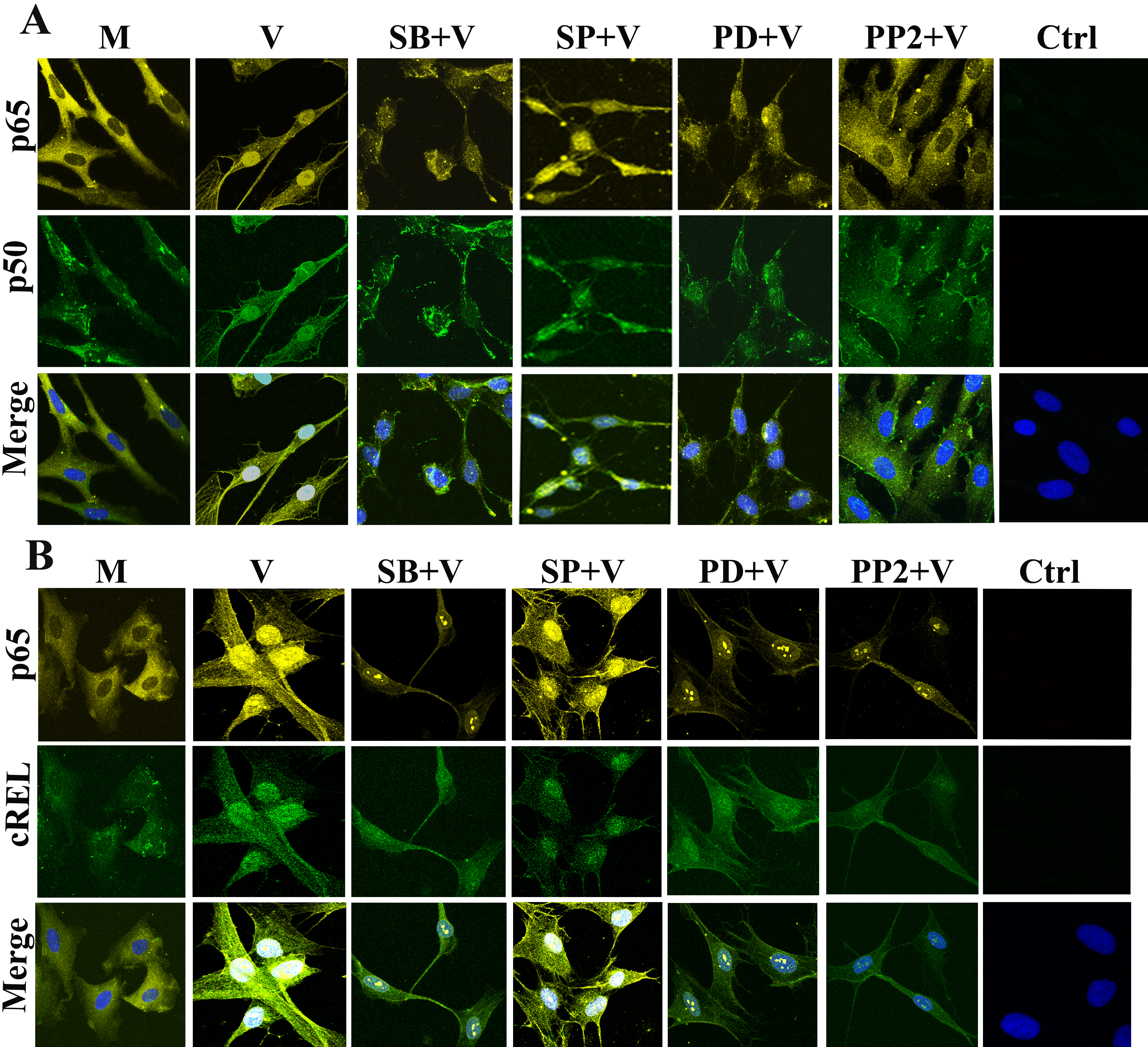
 Figure 3 of Rajaiya, Mol Vis 2009; 15:2879-2889.