Figure 1. Activation of NFκB subunits upon
adenoviral infection in keratocytes is shown. Whole cell extracts from
1 h post viral-infected (V) or mock-treated (M) cells, pretreated with
signaling inhibitors SB203580 (SB: p38), SP600125 (SP: JNK), PD98059
(PD: ERK), or PP2 (Src) were run on a 10% sodium dodecyl sulfate
polyacrylamide gel electrophoresis and immunoblotted using
anti-phospho-NFκB p65, NFκB p50, cREL, IKKα/β and IκB antibodies.
Viral-infected cells showed increased activation of NFκB p65, NFκB p50,
IKKα/β and IκB both at (A) 1 h and (B) 4 h after
infection. cREL phosphorylation was unchanged at 1 h, but increased at
4 h post infection. Actin levels to determine equivalent protein
loading are shown. Phosphorylation of p65, IKKα/β, and IκB was reduced
by SB, PD, and PP2 but SP only partially reduced the activation of NFκB
p65, IKKα/β, and IκB (A). At 4 h post infection, cREL
phosphorylation in viral-infected cells was reduced by all inhibitors
used (B).
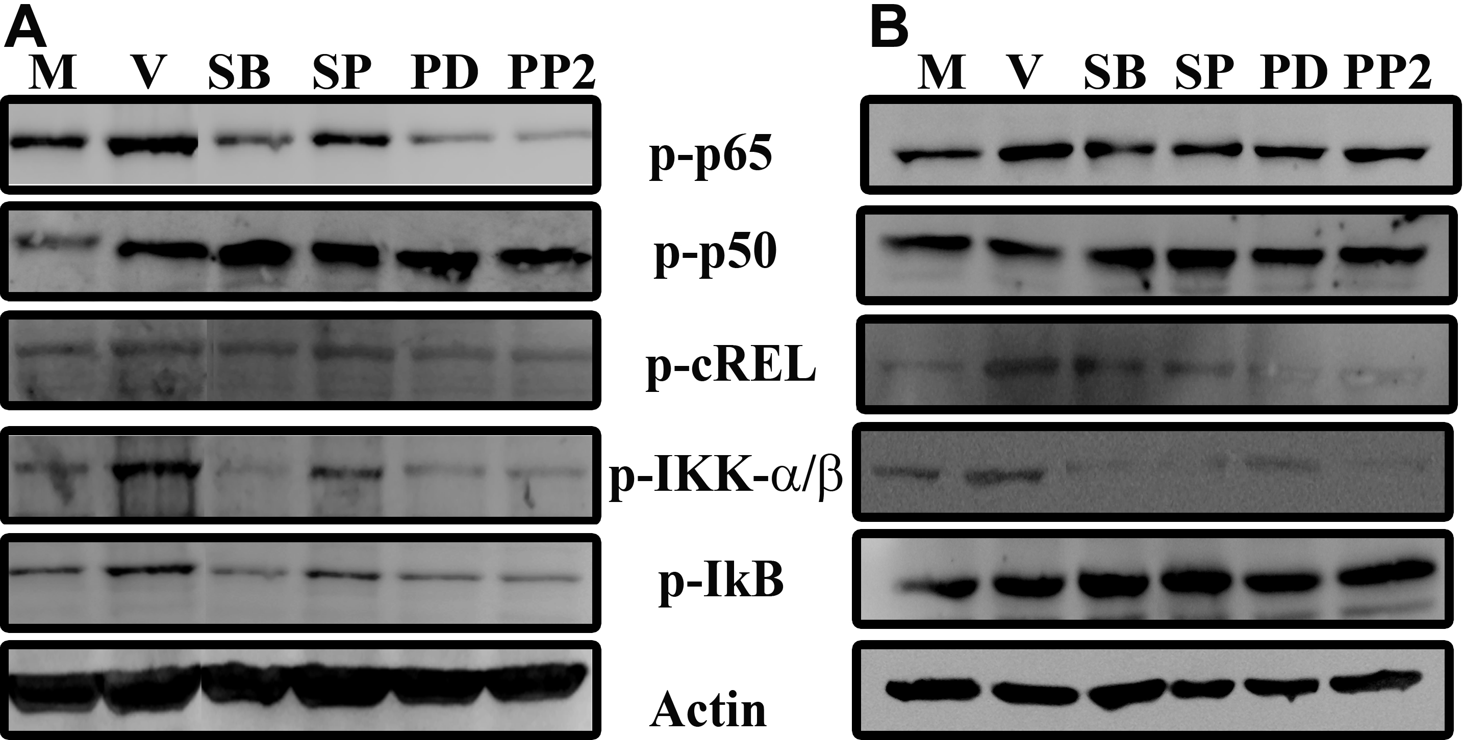
 Figure 1 of Rajaiya, Mol Vis 2009; 15:2879-2889.