Figure 2. Dopamine receptor deletion does
not alter photoreceptor cell survival in rd1 retinal organ
cultures. Retinas from D1−/−, rd1/+ (A),
D1+/−, rd1/rd1 (B), D1−/−,
rd1/rd1 (C), D2−/−, rd1/rd1 (D),
D4−/−, rd1/rd1 (E), and D5−/−,
rd1/rd1 (F) mice were harvested at postnatal day 2 and
grown in organ culture for 27 days in vitro. Wild-type retinas
maintained approximately five to six rows of cells in the ONL (A),
while the ONL of untreated rd1/rd1 cultures, regardless of DR
genotype, was reduced to approximately two to three rows (B-F).
Quantitative
analysis of photoreceptor survival as measured by ONL
thickness is shown in (G). Black bars represent the genotypes
shown in A-F and additional controls. Grey bars
represent organ cultures from D2−/−, D4−/−,
rd1/rd1 triple mutant mice treated with drugs as labeled. The
number of cultures is indicated on the column for each condition; error
bars indicate standard deviation. No significant difference is seen in
the ONL thickness among rd1 organ cultures, regardless of DR
genetic background or treatment. Similarly, no difference is seen among
rd1 heterozygous control organ cultures. Abbreviations: outer segments
(OS); inner segments (IS); outer nuclear layer (ONL); outer plexiform
layer (OPL); inner nuclear layer (INL); inner plexiform layer (IPL);
ganglion cell layer (GCL). The scale bar represents 10 μm.
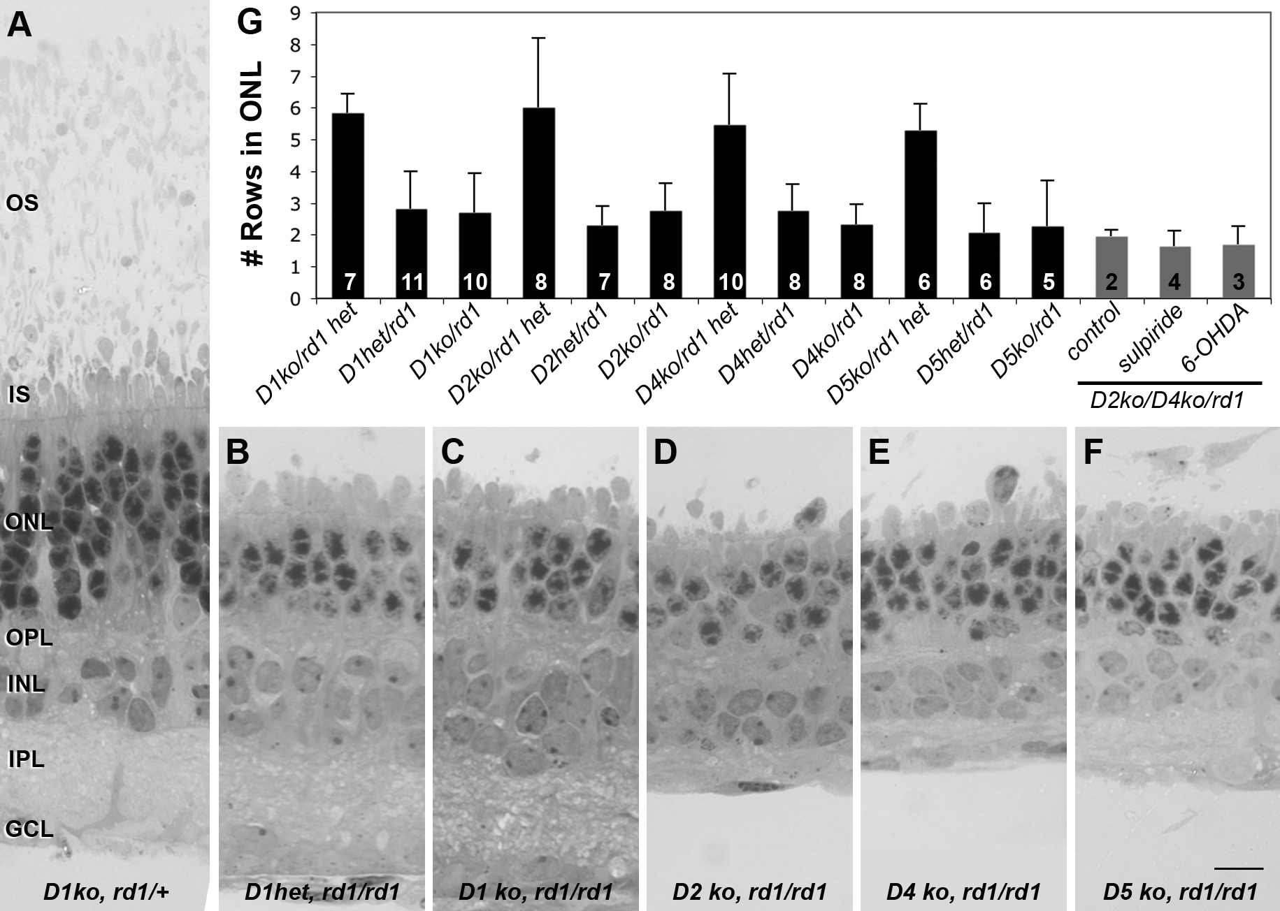
 Figure 2 of Ogilvie, Mol Vis 2009; 15:2868-2878.