Figure 1. A: Three different PCR–single stranded conformational analysis patterns on one gel, representing COL4A3 exons 17, 48, and 49. PCR fragments were loaded in succession from shortest to longest PCR fragment at 30-min intervals.
Exons 48 and 49 did not show any differences in elution shifts; in exon 17, the different patterns were subsequently sequenced.
B: Partial sequence of exon 17 with heterozygous substitution D326Y (976GT). C: Partial sequence of exon 17 with homozygous substitution 326Y (976TT). K1 to K4 marked patterns are patterns of three exons
(17, 48, and 49) of COL4A3 from keratoconus patients, multiplied by PCR and analysed with SSCA. C1 to C3 marked patterns are patterns of three exons
(17, 48, and 49) of COL4A3 from controls multiplied by PCR and analysed with SSCA. '1/1', '2/2' denote different genotypes at position 976 (1/2 being
GT and 2/2 being TT) in COL4A3. GG genotypes, being '1/1’ genotypes, are unmarked. Single stranded DNA (ssDNA) and double stranded DNA (dsDNA) patterns
are marked on the side of the SSCA gel.
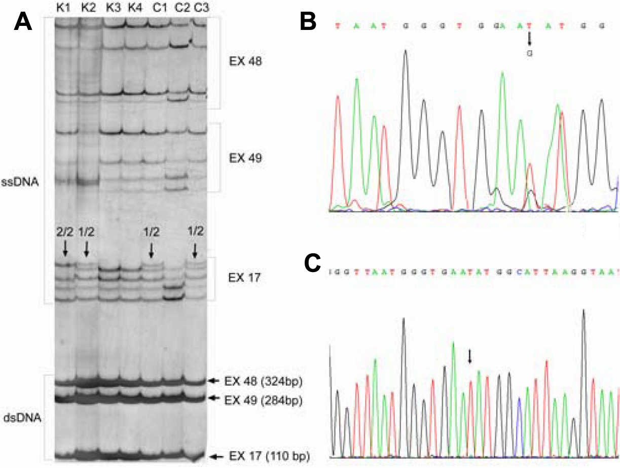
 Figure 1 of
Štabuc-Šilih, Mol Vis 2009; 15:2848-2860.
Figure 1 of
Štabuc-Šilih, Mol Vis 2009; 15:2848-2860.