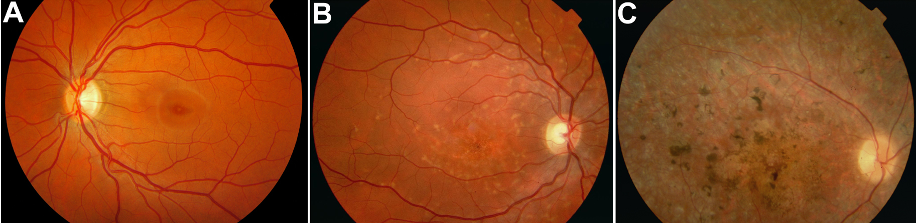
Figure 1. Fundus appearances according to
Fishman’s three-class system [
14].
A: This photograph represents fundus appearance type I, which is
characterized by a small atrophic-appearing foveal lesion. This lesion
can be surrounded by parafoveal or perifoveal yellowish white lesions.
B:
This
photograph
represents
fundus appearance type II, showing numerous
yellowish white fundus lesions extending beyond the vascular arcades.
C:
This
photograph
represents
fundus appearance type III, which is
characterized by extensive atrophic-appearing changes of the retinal
pigment epithelium throughout the posterior pole, extending beyond the
vascular arcades.
 Figure 1 of Ernest, Mol Vis 2009; 15:2841-2847.