Figure 4. The predicted secondary structures of the mutant and the wild-type amino acid sequences. The predicted secondary structures
of the wild-type amino acid sequence (A) and the mutant amino acid sequence (B) is shown. The target sequences are labeled by a red circle, which indicate that there is a helix in the wild type replaced
by a turn in the mutant type. Blue, helix; yellow, sheet; green, turn; black, coin.
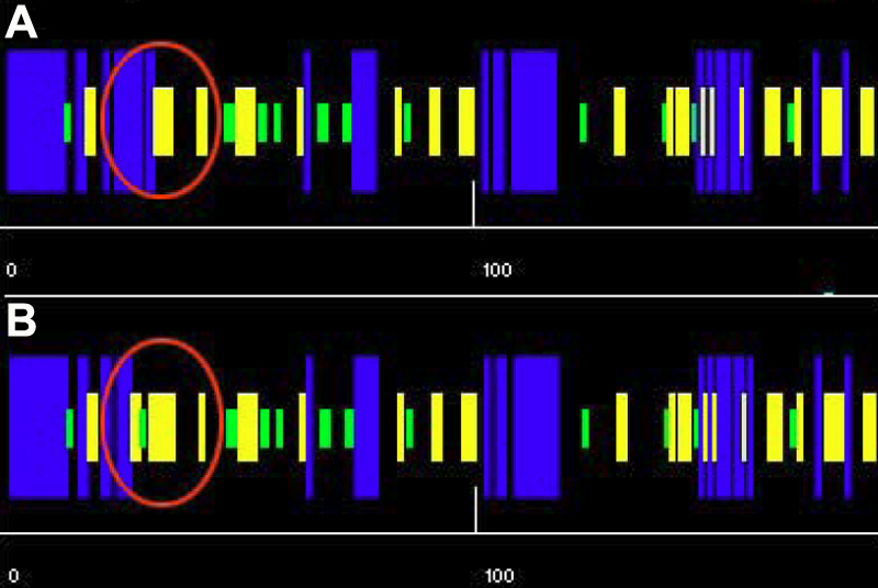
 Figure 4 of
Wang, Mol Vis 2009; 15:2813-2820.
Figure 4 of
Wang, Mol Vis 2009; 15:2813-2820.