Figure 2. The distribution in rat eyes of
intravitreally administered fluorescent dye labeled full-length IgG
(red). A and B show the distribution of the full-length
IgG in the live and post-mortem retina 6 h post intravitreal
administration, respectively. More antibodies were observed in the
post-mortem retina, compared to the live retina, suggesting the
presence of active elimination mechanisms for IgG in the live retina. A,
B: Cell nuclei were stained with DAPI (blue). Abbreviations:
inner limiting membrane (ILM), ganglion cell layer (GCL), inner
plexiform layer (IPL), inner nuclear layer (INL), outer plexiform layer
(OPL), outer nuclear layer (ONL), and external limiting membrane (ELM).
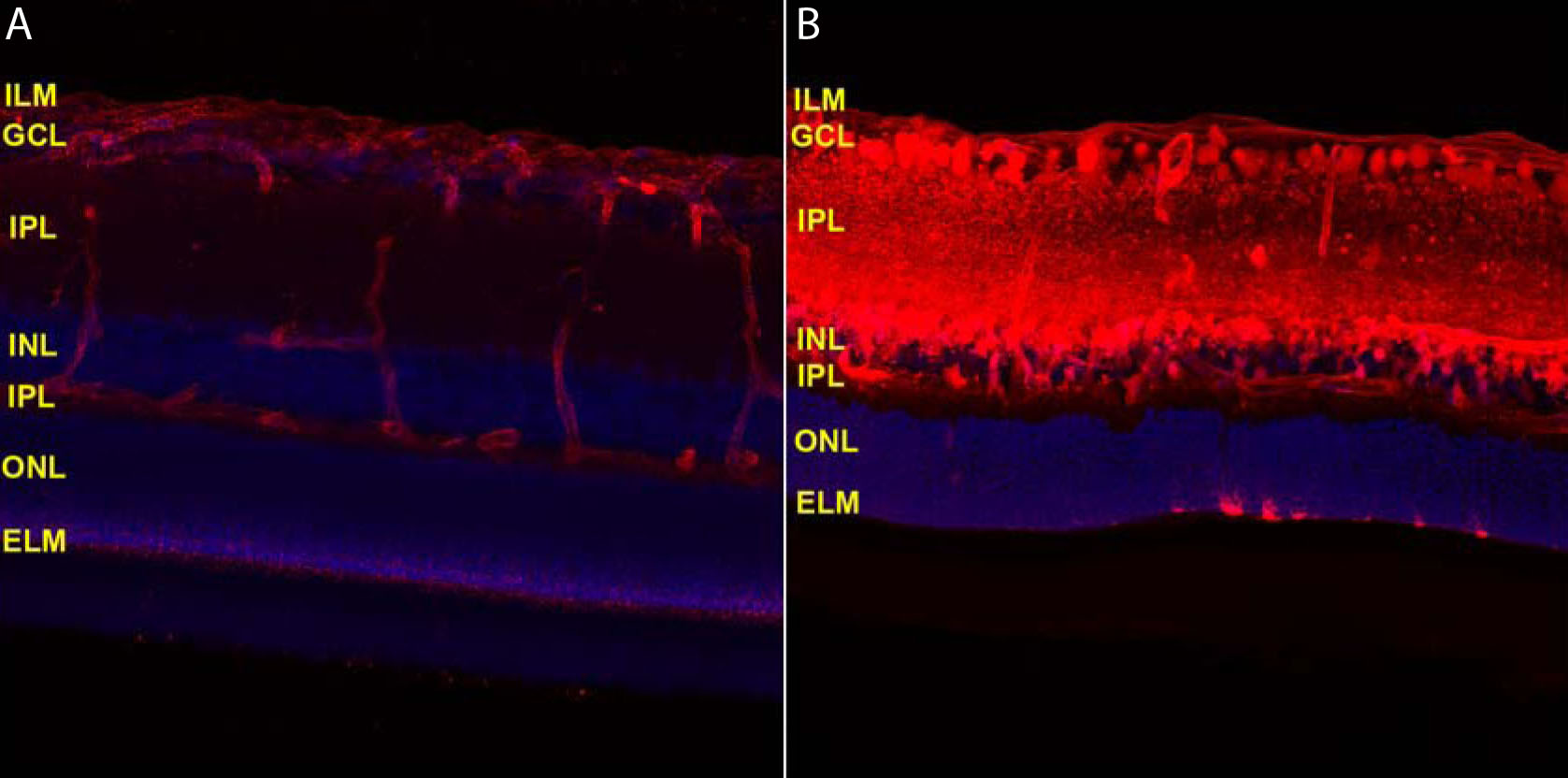
 Figure 2 of Kim, Mol Vis 2009; 15:2803-2812.