Figure 1. The distribution in rat eyes of
intravitreally administered fluorescent dye labeled full-length IgG
(red). A-C show the distribution of the full-length IgG
in the normal retina at 30 min, 6 h, and 15 h post intravitreal
administration, respectively. D and E show the
distribution of the full-length IgG in the laser-photocoagulated retina
at 1 h and 3 h post intravitreal administration, respectively.
Intravitreally administered full-length antibody overcame the inner
limiting membrane barrier and diffused into the deeper retinal
structures in both laser photocoagulated and control rat retinas. A–E:
Cell
nuclei were stained with DAPI (blue). Abbreviations: inner
limiting membrane (ILM), ganglion cell layer (GCL), inner plexiform
layer (IPL), inner nuclear layer (INL), outer plexiform layer (OPL),
outer nuclear layer (ONL), external limiting membrane (ELM), and
Bruch’s membrane (BM).
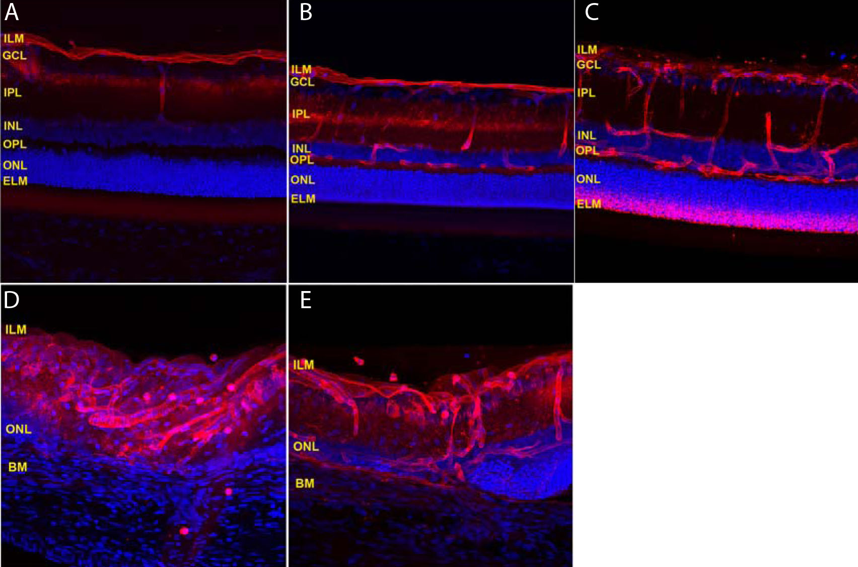
 Figure 1 of Kim, Mol Vis 2009; 15:2803-2812.