Figure 2. Fluorescence microscopy controls. Retinal cross-sections counterstained with DAPI (A) without the primary anti-green fluorescent protein (GFP) antibody that were counterstained with the secondary antibody showed
no GFP labeling (B). DAPI stained RGC layer (C) from specimen double-labeled for GFP (D) and Thy1.2 are shown (E). The merged image of panels C-E shows Thy1.2 RGCs without GFP positivity (F). Abbreviations: ONL represents outer nuclear layer, INL represents inner nuclear layer, RGC represents retinal ganglion
cell. Scale bar equals to 50 µm.
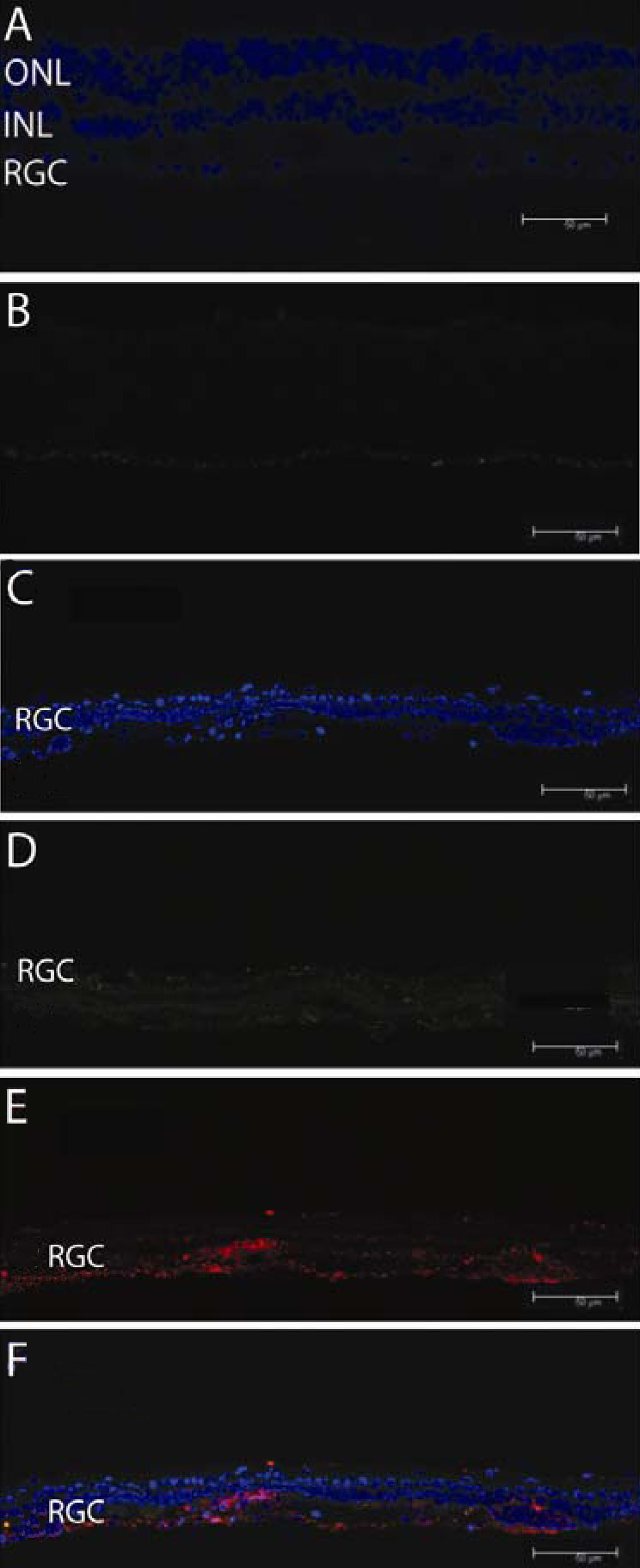
 Figure 2 of
Koilkonda, Mol Vis 2009; 15:2796-2802.
Figure 2 of
Koilkonda, Mol Vis 2009; 15:2796-2802.