Figure 1. Fluorescence Microscopy GFP.
Retinal flat mounts inoculated with scAAV-GFP revealed some green
fluorescent protein (GFP) positive cells (A). Confocal
microscopy revealed many more GFP positive cells by immunofluorescence (B).
RGCs
labeled with the Brn3a antibody (C) and merged images
revealed GFP expressing cells co-localized exclusively with those
expressing Brn3a (D). Higher power confocal micrographs of
scAAV-GFP injected eyes show nuclei labeled by DAPI (E). Cells
expressing GFP (F) or Thy1.2 (G) co-localized (H).
Cryo-sectioned retinas of scAAV-GFP injected eyes counterstained with
DAPI revealed the nuclei of cells in the retinal ganglion cell layer
(RGC), inner nuclear layer (INL) or outer nuclear layer (ONL; I).
GFP
immunofluorescence is seen exclusively in the ganglion cell layer (J)
whose
RGCs were labeled by Thy1.2 (K). Colocalization of GFP and
Thy1.2 cells is shown in the merged panel (L).
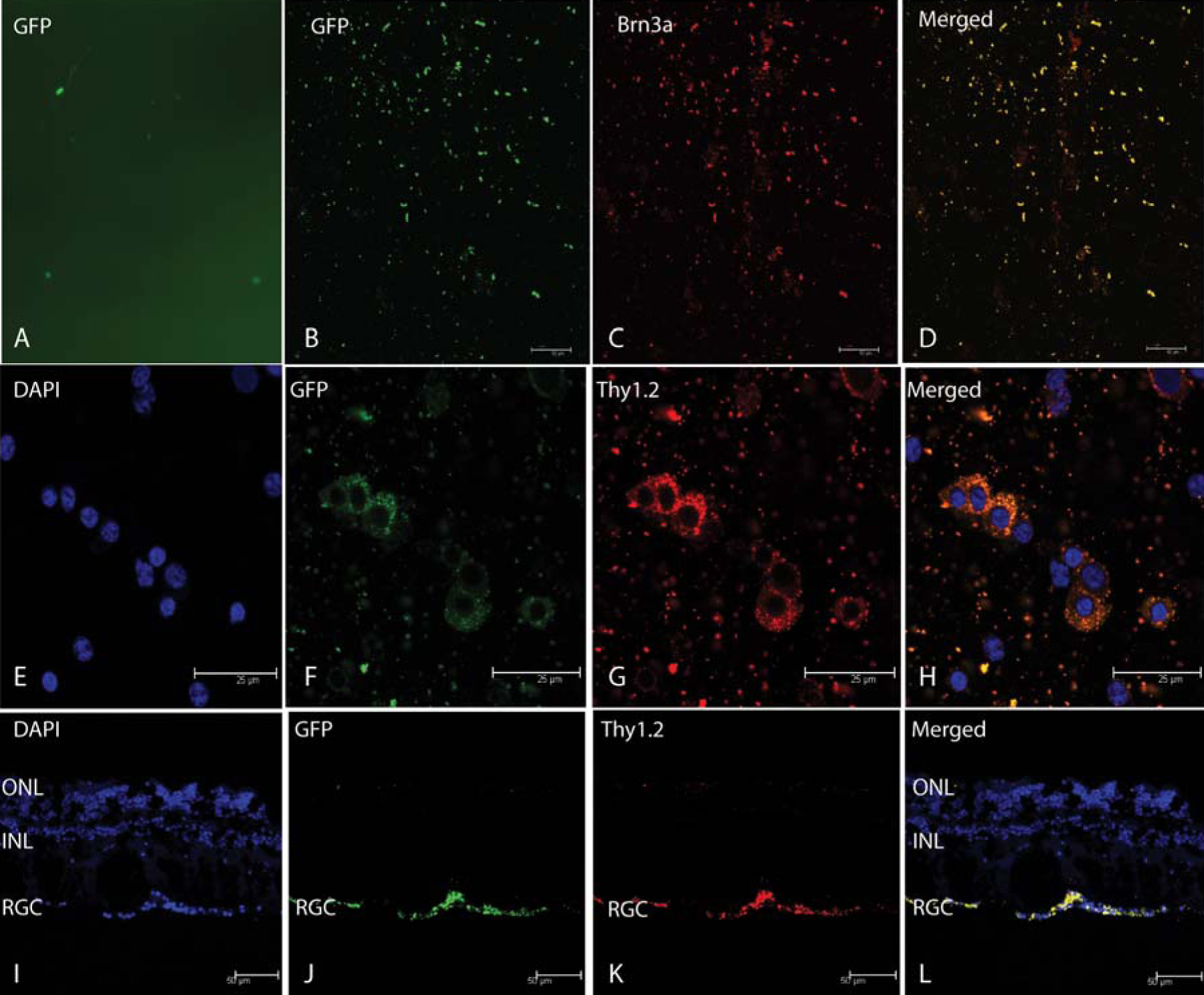
 Figure 1 of Koilkonda, Mol Vis 2009; 15:2796-2802.