Figure 4. Histological results at 1 year.
Histological corneal sections of treated animals (A–D)
show an implanted corneal epithelium of a multilayer configuration with
maturation from polygonal basal cells to flat superficial cells.
Beneath the epithelium there is an acellular colagenous hyaline layer
(arrows) with isolated keratocytes. The median and deep stroma as well
as the Descemet membrane and endothelium have a normal appearance
(hematoxylin and eosin stain; A). Periodic acid-Schiff
stain shows epithelial-like cells with clear cytoplasm at the basal
layer of the corneal epithelium with a continuous basement membrane. A
slightly periodic acid-Schiff positive acellular layer is seen between
the epithelium and the normal corneal stroma (arrows; B). At
the central area of the cornea, besides a thin homogenous layer beneath
the epithelium, the remaining corneal stroma disclosed a normal
lamellar configuration with interspread keratocytes (Masson stain; C).
In the peripheral cornea, a thick layer of proliferated myofibroblasts
and colagenous disposition deposits containing a few inflammatory cells
is present between the implanted epithelium and the remaining corneal
stroma (Masson stain; D). In the control group (E–H)
an irregular regenerative epithelium from the limbal conjunctiva can be
distinguished over the remaining stroma and a subepithelial scarring
with inflammatory cells and neovascularization is present (Hematoxylin
and eosin stain; E). A taller epithelium compared to
normal/treated corneas is evident, with incomplete surface cell
differentiation (Hematoxylin and eosin stain; F). Periodic
acid-Schiff of the peripheral cornea discloses the presence of goblet
cells within the corneal epithelium and a scarring of the underlying
corneal stroma (G). Central cornea of a control eye shows a
multilayered squamous-like corneal epithelium and a thick layer of
active scar containing myofibroblasts, colagenous deposits, and
inflammatory cells at the interface within the remaining corneal stroma
(Masson stain; H).
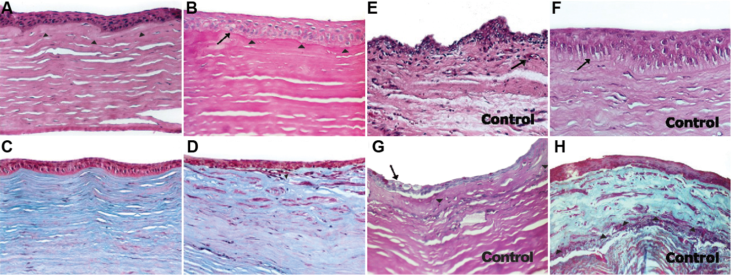
 Figure 4 of Gimeno, Mol Vis 2009; 15:2771-2779.