Figure 2. Clinical results at 1 year after
surgery. Five eyes of the treated group displayed avascular and
re-epithelialized corneas, with no signs of limbal stem cell deficiency
(B, C, E, F, and G), one animal
showed corneal neovascularization (A), one stayed with a stromal
leucoma (D) and the other suffered from neovascularization that
regressed thereafter leaving a stromal leucoma (H). Figure I
shows one eye of the control group with no implant, at 3 months. The 3
animals of the control group implanted with PPP showed signs of limbal
stem cell deficiency: one rabbit had to be sacrificed at 120 days
because of a severe infection due to a IV grade corneal abscess (J),
another spontaneously died at 240 days showing a small inferior leucoma
and epithelial defects (K) and the remaining rabbit displayed
total irregularity of the corneal surface at one year after surgery (L).
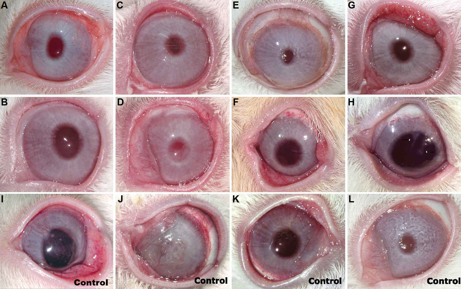
 Figure 2 of Gimeno, Mol Vis 2009; 15:2771-2779.