Figure 1. Allograft of corneal epithelial
cell sheet on autologous platelet poor plasma clot. A, B,
D: Once the lamellar keratectomy is performed, the rabbit
stromal surface is covered with a corneal epithelial cell sheet, which
is sutured with eight noncontinuous 10.0 nylon sutures. C: The
corneal epithelial cell sheet presents a stratified epithelium
consisting of four layers, inferior cells are polygonal and disclose a
prominent nucleolus while cells of mid epithelium show irregular
morphology, and superficial cells are flattened with a compact ovoid
nucleus of homogeneous chromatin.
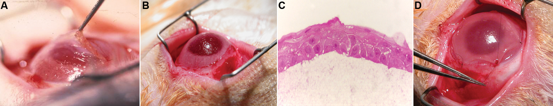
 Figure 1 of Gimeno, Mol Vis 2009; 15:2771-2779.