Figure 2. This shows changes in expression of RSK2; a portion of 2D-gel that includes the protein spot of RSK2 (arrow) is shown at high
magnification. A: Total lysates of tissue sample from failed filtering blebs (Failed bleb) and from Tenon obtained during cataract surgery
(Control) were applied to 12% polyacrylamide gel (SDS PAGE), and blotted with anti-RSK2 antibody. Migration position of RSK2
is indicated. B: The panel on the left shows RSK2 expression in Tenon’s capsule derived from cataract surgery stained with an anti-RSK2 antibody.
The right-hand panel is stained without the primary antibody, as a negative control. Arrows show the actual protein spots,
identified as RSK2.
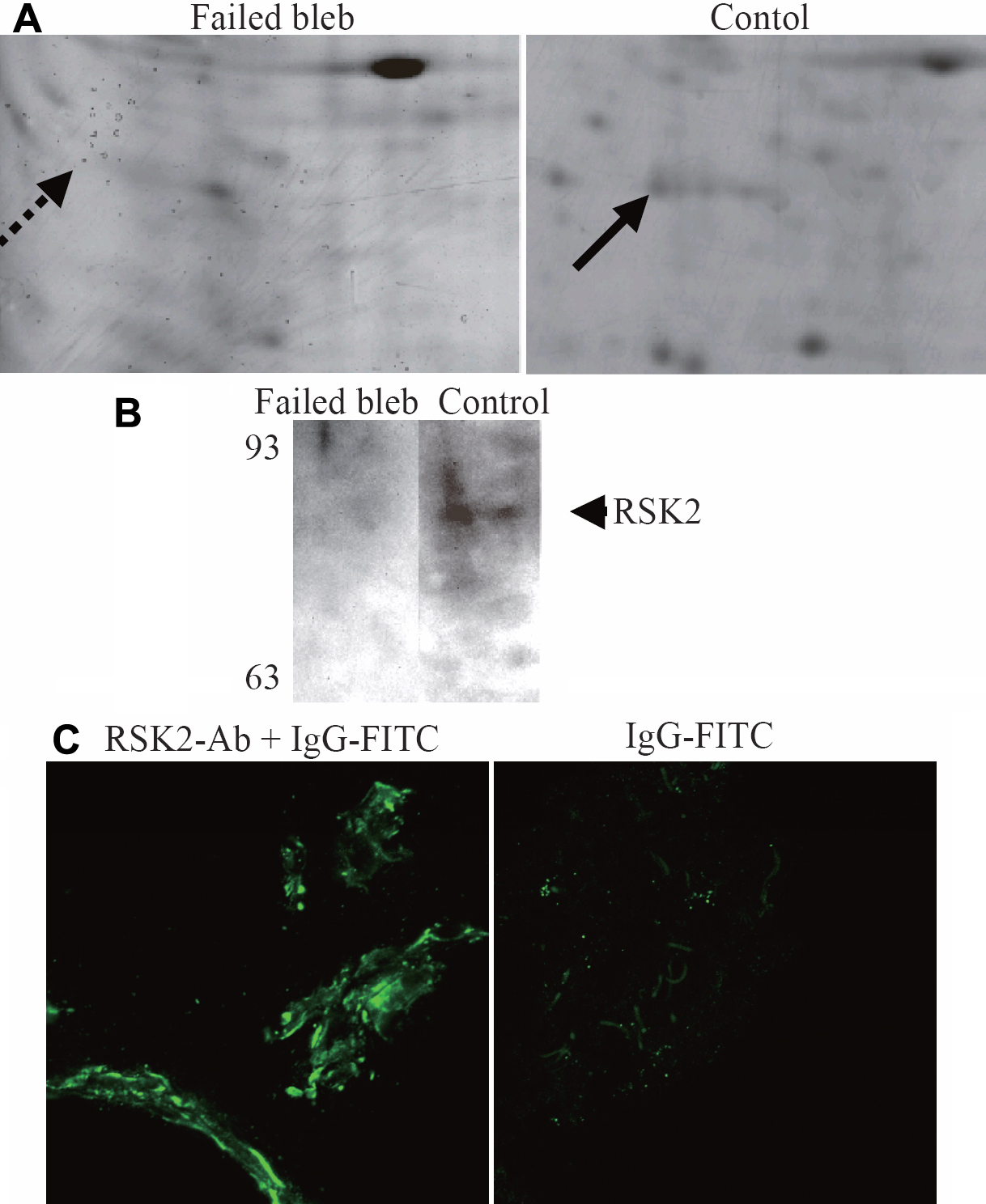
 Figure 2 of
Kanamoto, Mol Vis 2009; 15:2762-2770.
Figure 2 of
Kanamoto, Mol Vis 2009; 15:2762-2770.