Figure 2. Development of posterior
capsular opacity in murine IOL-implanted eyes. Representative
micrographs of murine IOL-implanted eyes, stained with hematoxylin and
eosin on post-operative day 1 (A) or day 5 (B) and
high-magnification images of the posterior lens capsule (C, D;
square area marked in A and B, respectively). The
arrows depict lens epithelial cells migrating underneath the posterior
capsule on post-operative day 5 (D), whereas no cells were seen
on post-operative day 1 (C). The bar shown in A
represents 500 µm and applies for (B) as well. The bar shown in C
represents 200 µm and applies for (D) as well.
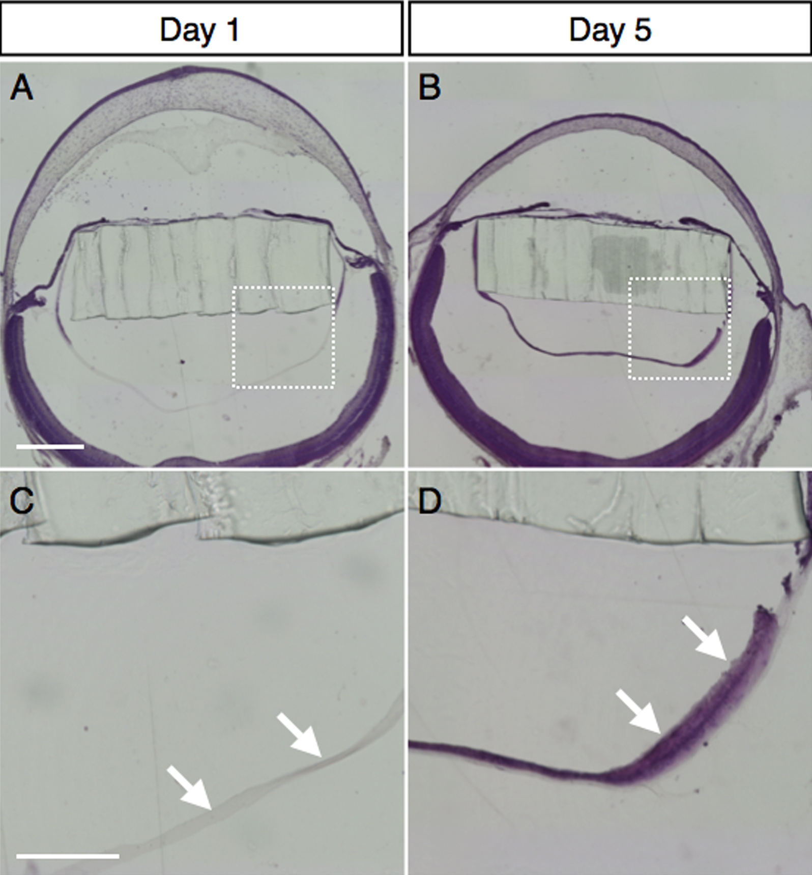
 Figure 2 of Kurihara, Mol Vis 2009; 15:2751-2761.