Figure 1. Extra-capsular crystalline lens
extraction and IOL implantation in murine eyes. A-C:
Corneal incision with a surgical knife and Vannas scissors. D:
Continuous curvilinear capsulorrhexis with forceps. E: Lens
extraction with hydrodissection. F: Inflation of the capsular
bag with a viscoelastic substance. G: Fabricated IOL buttons (2
mm diameter). H, I: IOL insertion. J, K:
Removal of the viscoelastic substance and closure of the corneal wound
with interrupted 11–0 nylon sutures.
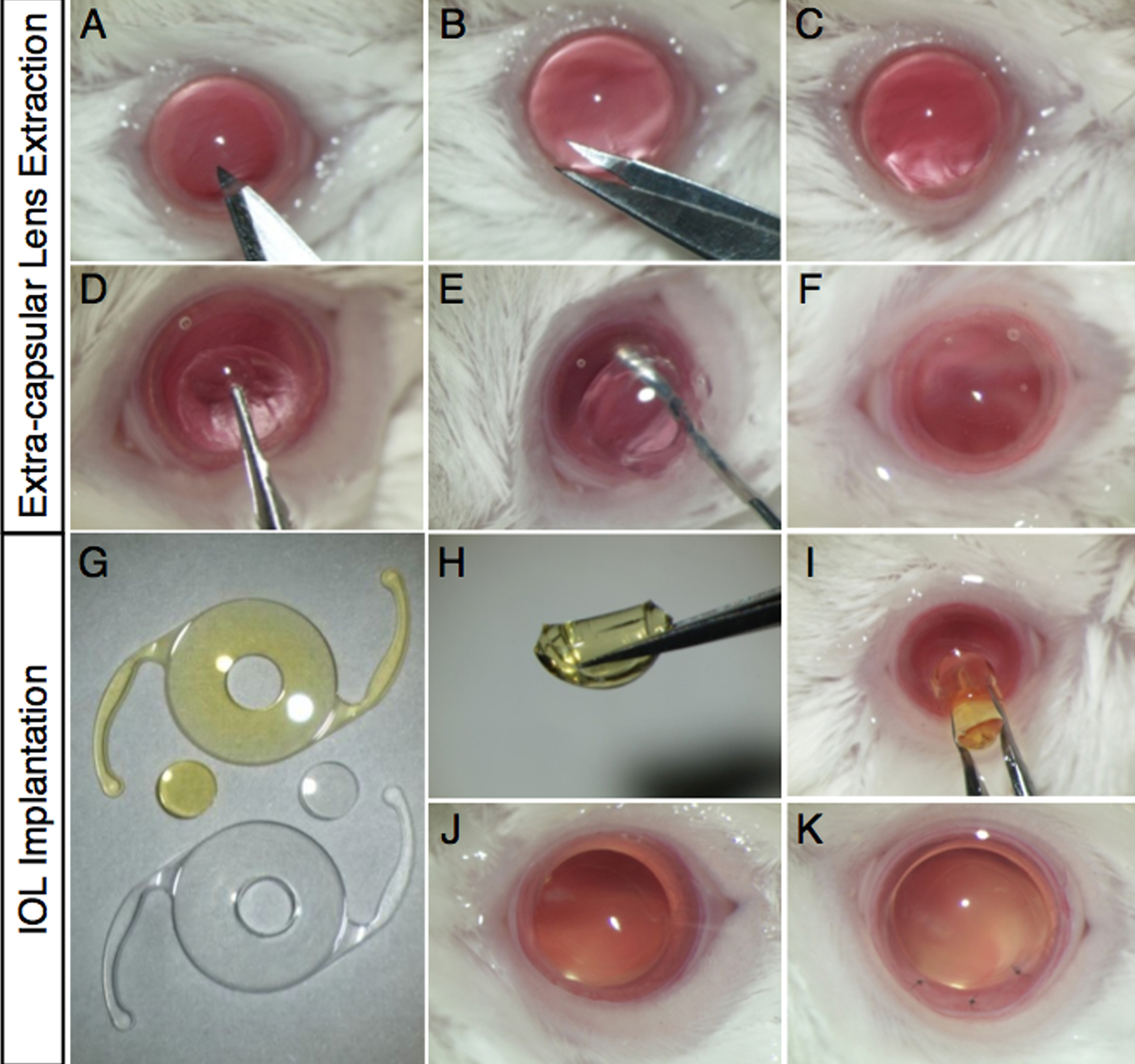
 Figure 1 of Kurihara, Mol Vis 2009; 15:2751-2761.