Figure 6. Electron microscopy of
HESC-derived RPE cells after exposure to human retinal explant in
vitro. A: Control HESC-derived RPE cells, which were not
exposed to human retina, have apical microvilli (AMv) and contain
melanin granules (white arrows). B: HESC-derived RPE exposed to
the photoreceptor surface of human retina for 48 h appear more mature.
Note the close association of the RPE apical microvilli with the
photoreceptor outer segment (OS), the abundance of pigmented melanin
granules within the apical region of the cell, the nucleus (N), and the
numerous lipid deposits (L) located toward the basal portion of the
cell. C: Apical microvilli surround a human photoreceptor outer
segment. D: An outer segment is engulfed by the HESC-derived
RPE cell. E: A high magnification image of lipid deposits (L)
observed in the basal portion of a HESC-derived RPE after exposure to
human retina. The formation of lipid deposits is indicative of the end
stages of phagocytosis. Several features associated with RPE cell
function are present in the cells including (F) tight junctions
(black arrow) and coated pits (red box, indicated with arrows at higher
magnification
in G). Cells also contain a high number of coated vesicles
within the apical portion of the cell (H) and develop basal end
feet and infolding of the basal membrane (I). Scale bars equal 2
μm in A-C, 1 μm in D-F,I, and 500
nm in G,H.
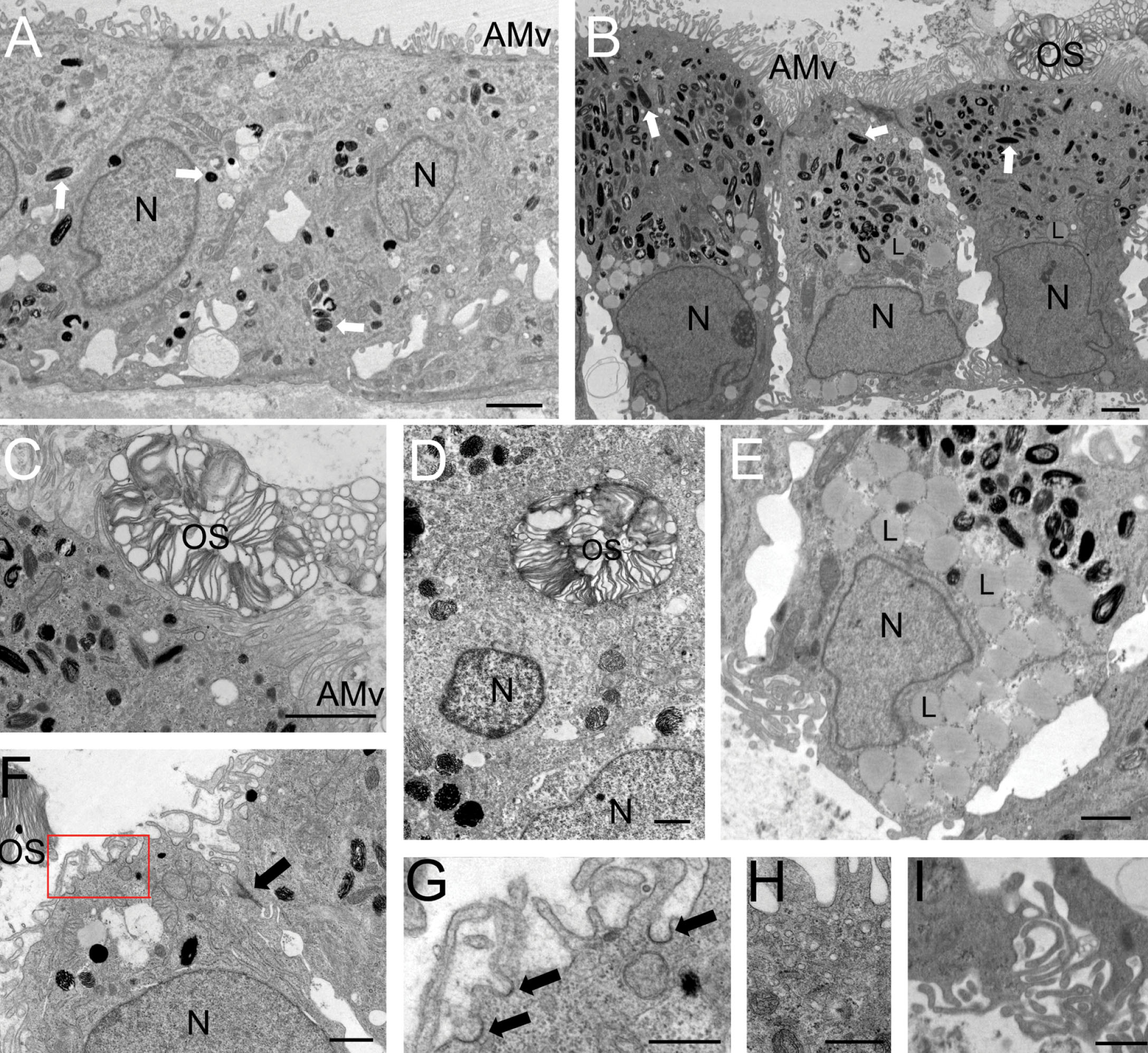
 Figure 6 of Carr, Mol Vis 2009; 15:283-295.